Copyright
©The Author(s) 2020.
World J Radiol. Sep 28, 2020; 12(9): 195-203
Published online Sep 28, 2020. doi: 10.4329/wjr.v12.i9.195
Published online Sep 28, 2020. doi: 10.4329/wjr.v12.i9.195
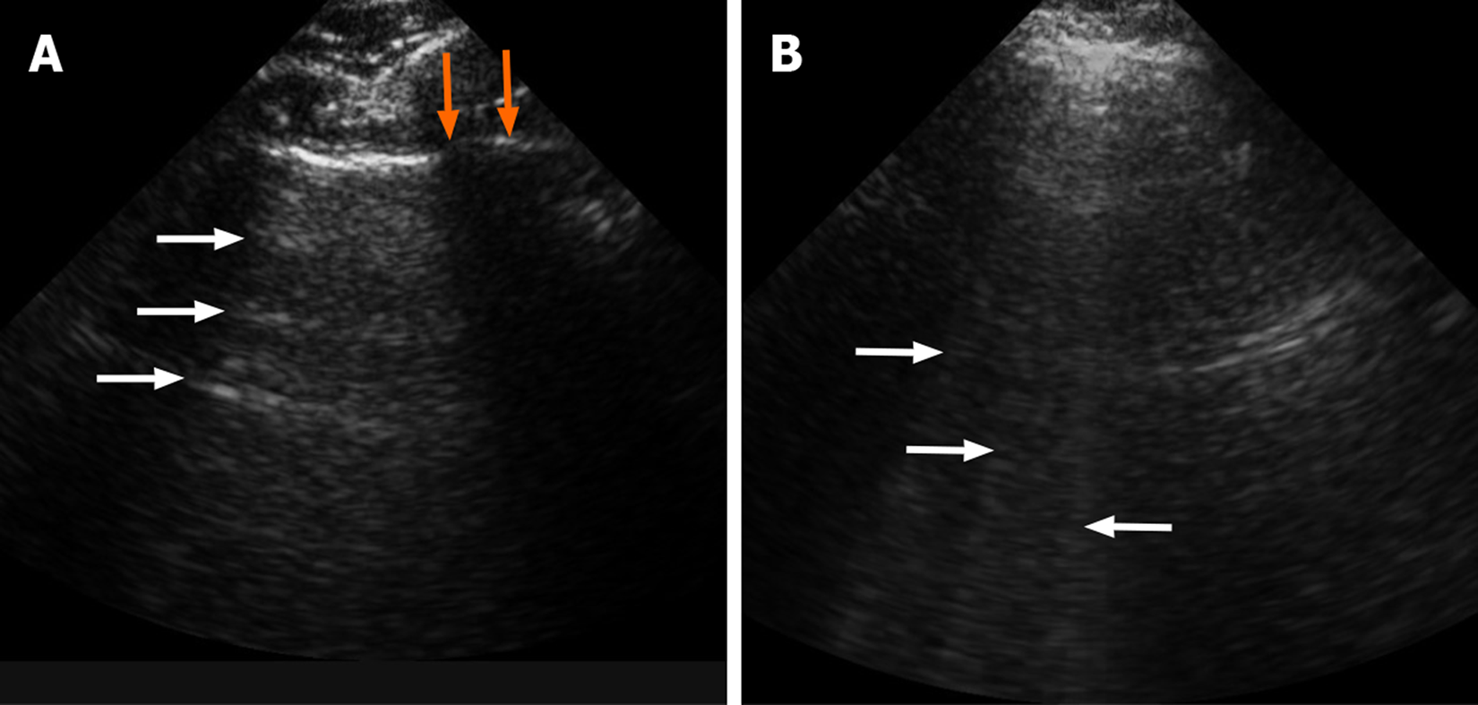
Figure 6 Ultrasonography imaging of the lung ultrasound of the normal lung shows horizontal artifacts, so called “A” lines (white arrows).
(A) and vertical heterogenous defect or so called vertical “B” lines in case of patient with consolidation and/or atelectasis (B). Also note normal rib shadow in figure A (orange arrows).
- Citation: Gandhi D, Ahuja K, Grover H, Sharma P, Solanki S, Gupta N, Patel L. Review of X-ray and computed tomography scan findings with a promising role of point of care ultrasound in COVID-19 pandemic. World J Radiol 2020; 12(9): 195-203
- URL: https://www.wjgnet.com/1949-8470/full/v12/i9/195.htm
- DOI: https://dx.doi.org/10.4329/wjr.v12.i9.195


