Copyright
©The Author(s) 2020.
World J Radiol. Aug 28, 2020; 12(8): 172-183
Published online Aug 28, 2020. doi: 10.4329/wjr.v12.i8.172
Published online Aug 28, 2020. doi: 10.4329/wjr.v12.i8.172
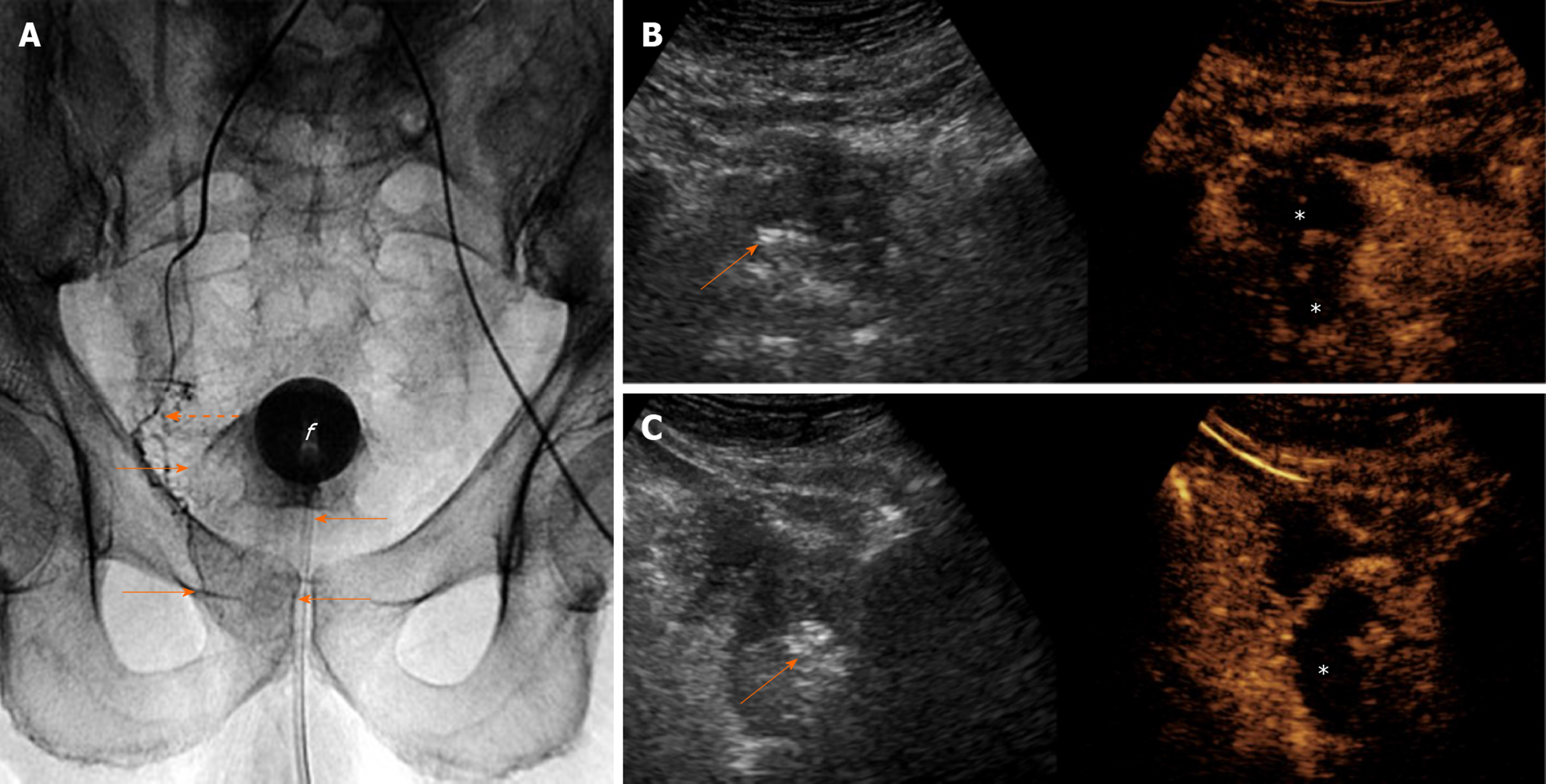
Figure 5 Intraprocedural sonographic evaluation of prostatic artery embolization with i.
v.-contrast-enhanced ultrasonography. A: Digital radiograph (anteroposterior projection) immediately post-right prostatic artery embolization shows the tip of the microcatheter (dotted arrow) at the proximal (extraprostatic) part of the right prostatic artery. Pooling of the contrast of the embolic mixture has caused opacification of the right hemiprostate (arrows). “f” indicates the balloon of the Foley catheter; B and C: Transabdominal contrast-enhanced ultrasonography (CEUS), axial and right parasagittal section, respectively, with standard (i.v.) injection of the echo-enhancer, confirms adequate devascularization (infarction) of the right hemiprostate (asterisks). Findings are presented in a “split-screen” mode with unenhanced, reference B-mode image on the left, and corresponding CEUS image on the right. Stippled and linear hyperechogenicities appear in the right hemiprostate. They are more striking (arrows) on the unenhanced, reference B-mode images.
- Citation: Moschouris H, Dimakis A, Anagnostopoulou A, Stamatiou K, Malagari K. Sonographic evaluation of prostatic artery embolization: Far beyond size measurements. World J Radiol 2020; 12(8): 172-183
- URL: https://www.wjgnet.com/1949-8470/full/v12/i8/172.htm
- DOI: https://dx.doi.org/10.4329/wjr.v12.i8.172


