Copyright
©The Author(s) 2020.
World J Radiol. Aug 28, 2020; 12(8): 172-183
Published online Aug 28, 2020. doi: 10.4329/wjr.v12.i8.172
Published online Aug 28, 2020. doi: 10.4329/wjr.v12.i8.172
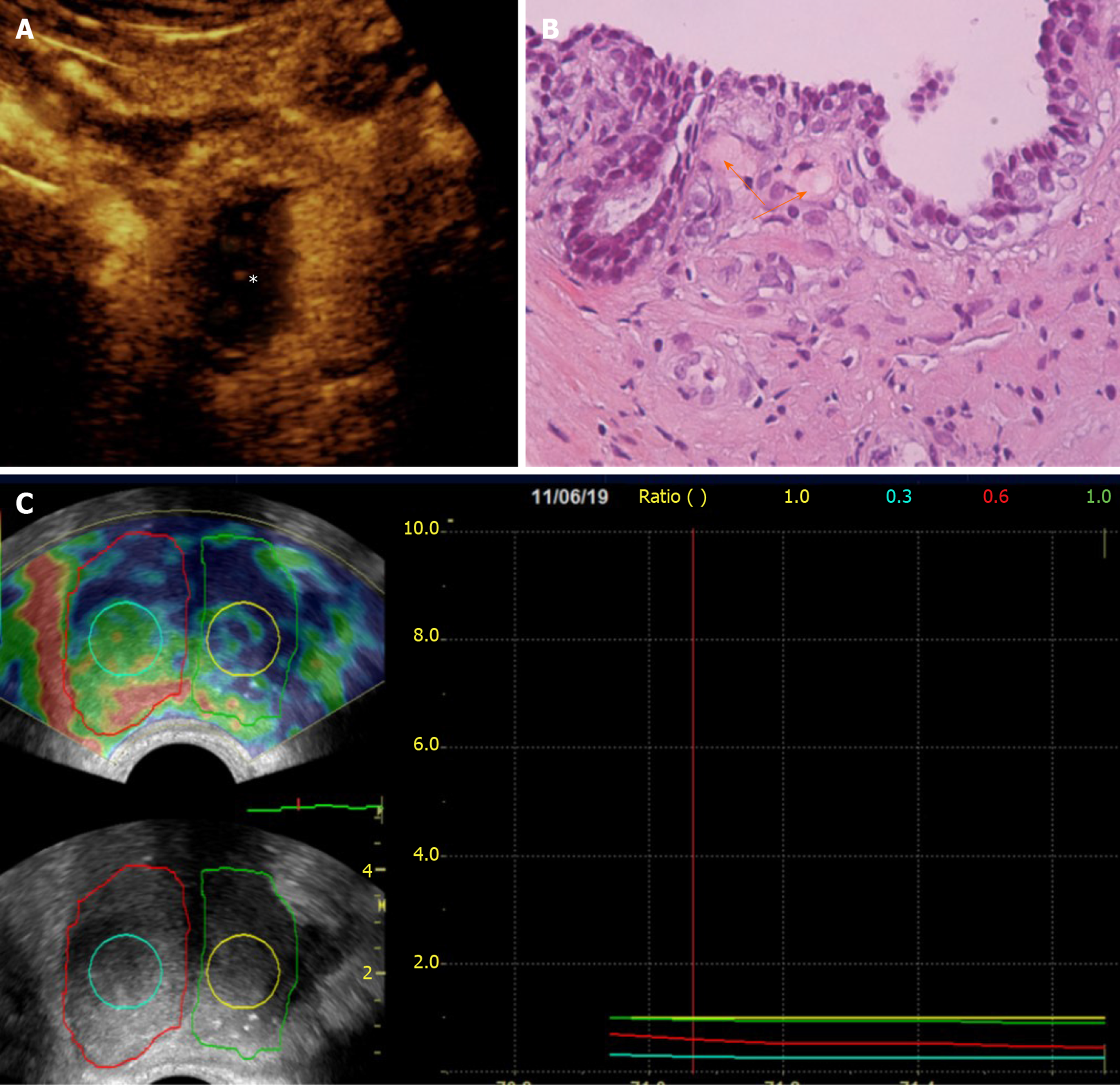
Figure 4 Correlation of contrast-enhanced ultrasonography, histopathologic and elastographic findings post-unilateral (right) prostatic artery embolization.
A: Transabdominal contrast-enhanced ultrasonography (CEUS) image (axial section) obtained one week post-prostatic artery embolization (PAE) shows extensive infarction of the right hemiprostate (asterisk); B: Histopathology (Hematoxylin-Eosin stain, original magnification × 40) of a transrectal US-guided biopsy specimen of the right transitional zone confirms the presence of eosinophilic microspheres (arrows) in arterioles surrounding a prostatic acinus. This biopsy had been performed 12 mo post-PAE, for recently elevated prostate-specific antigen and was negative for cancer; C: Transrectal strain elastographic images (axial sections) of the prostate 13 mo post-PAE shows increased strain in the right hemiprostate and in the center of the right transitional zone (outlined by the red and turquoise line, respectively) compared to the left hemiprostate and to the center of the left transitional zone (outlined by the green and yellow line, respectively). In this elastogram, blue color tones indicate hard (stiffer) tissues, red indicate soft tissues, and yellow or green tones indicate tissues of intermediate stiffness. This patient experienced significant and long-lasting symptomatic improvement post-PAE (75% reduction of the International Prostate Symptom Score, 2 years post-PAE).
- Citation: Moschouris H, Dimakis A, Anagnostopoulou A, Stamatiou K, Malagari K. Sonographic evaluation of prostatic artery embolization: Far beyond size measurements. World J Radiol 2020; 12(8): 172-183
- URL: https://www.wjgnet.com/1949-8470/full/v12/i8/172.htm
- DOI: https://dx.doi.org/10.4329/wjr.v12.i8.172


