Copyright
©The Author(s) 2020.
World J Radiol. Aug 28, 2020; 12(8): 172-183
Published online Aug 28, 2020. doi: 10.4329/wjr.v12.i8.172
Published online Aug 28, 2020. doi: 10.4329/wjr.v12.i8.172
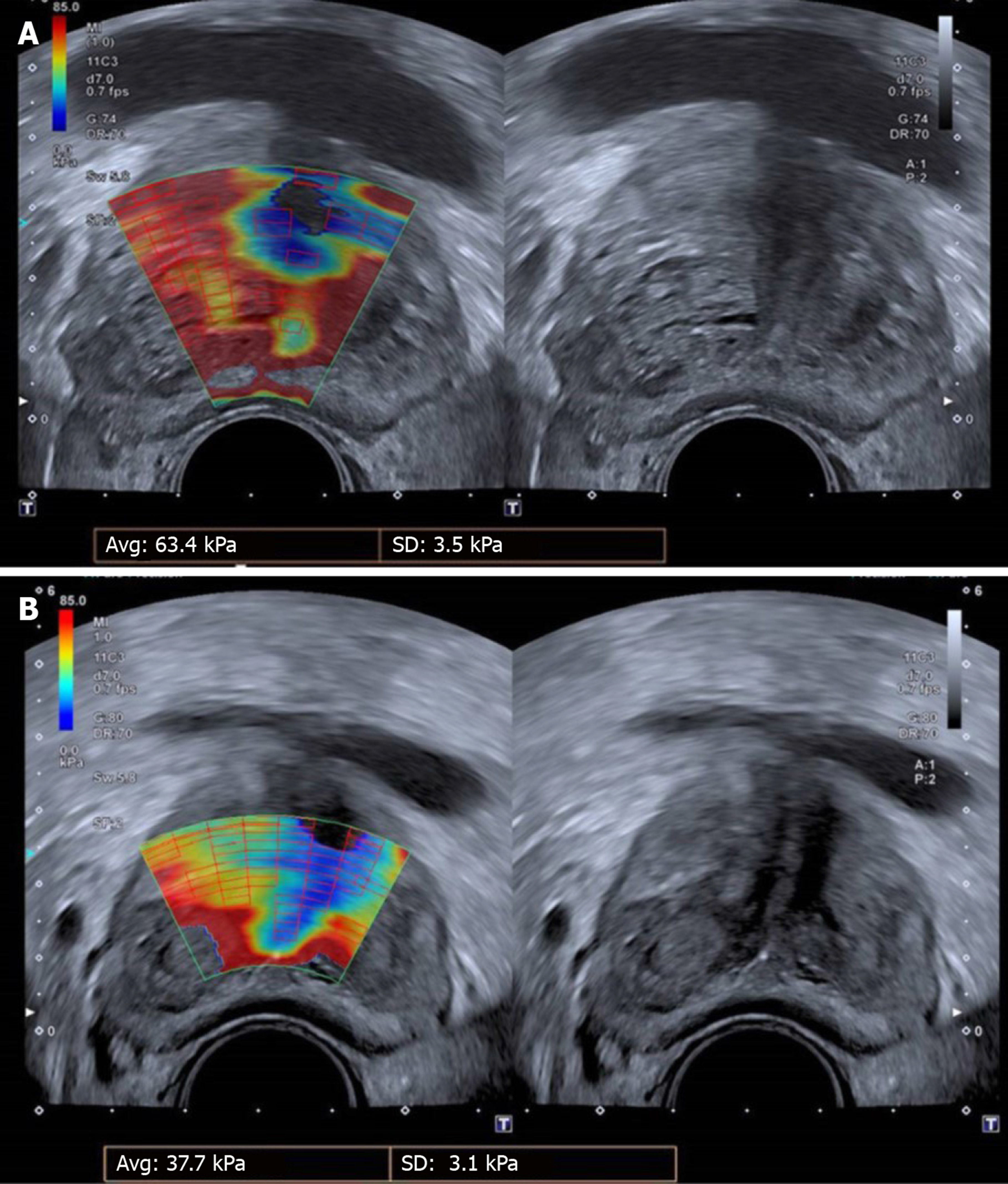
Figure 3 Shear-wave elastography of the prostate.
A: Before; B: Three months post-bilateral prostatic artery embolization (PAE). A significant reduction (40.5%) in the elastic modulus (EM) of the transitional zone is demonstrated, indicating increased prostatic tissue elasticity post-PAE. The elastographic changes were accompanied by marked improvement of clinical parameters (96.7% reduction of the International Prostate Symptom Score, 100% reduction of quality of life) and of prostate volume (55% reduction) and post-void residual volume (76% reduction). In this examination, red color tones indicate hard (stiffer) tissues, with high EM values (in kPa); blue tones indicate soft tissues, with low EM values; yellow or green tones indicate tissues of intermediate stiffness. Case courtesy of Dr. de Assis AM, Interventional Radiology Department, Radiology Institute, University of Sao Paulo Medical School.
- Citation: Moschouris H, Dimakis A, Anagnostopoulou A, Stamatiou K, Malagari K. Sonographic evaluation of prostatic artery embolization: Far beyond size measurements. World J Radiol 2020; 12(8): 172-183
- URL: https://www.wjgnet.com/1949-8470/full/v12/i8/172.htm
- DOI: https://dx.doi.org/10.4329/wjr.v12.i8.172


