Copyright
©The Author(s) 2020.
World J Radiol. Aug 28, 2020; 12(8): 172-183
Published online Aug 28, 2020. doi: 10.4329/wjr.v12.i8.172
Published online Aug 28, 2020. doi: 10.4329/wjr.v12.i8.172
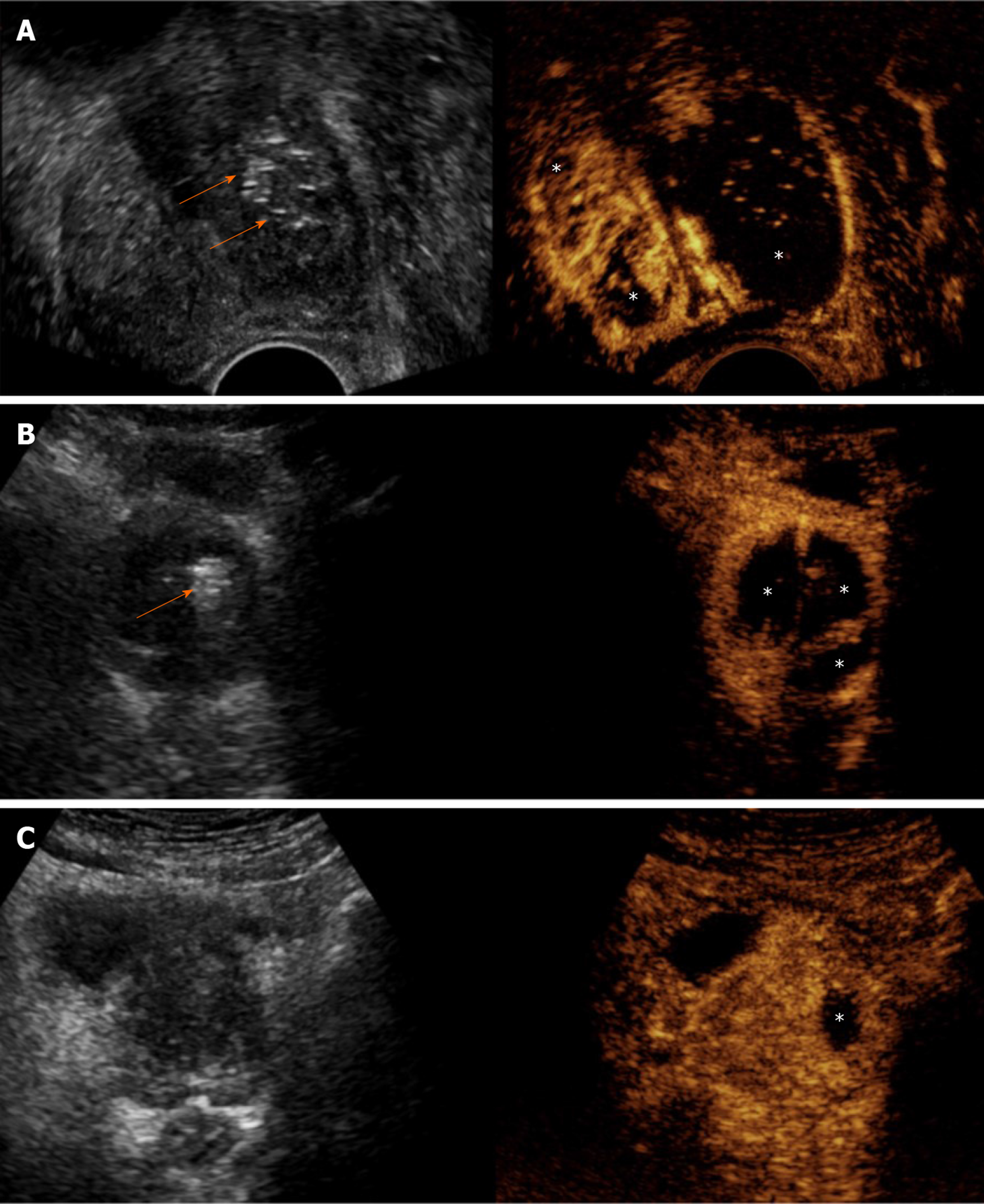
Figure 2 Varying extent and distribution of prostatic infarcts in patients with different clinical outcomes.
Findings are presented in a “split-screen” mode with unenhanced, reference B-mode image on the left, and corresponding contrast-enhanced ultrasonography (CEUS) image on the right. A: Transrectal CEUS 5 d post-bilateral prostatic artery embolization (PAE) in a patient with indwelling bladder catheter shows extensive infarction of the left prostatic lobe and smaller infarcts in the right. Removal of the Foley catheter and spontaneous voiding was possible a week later; B: Transabdominal CEUS (t.a.-CEUS) one day post-bilateral PAE shows extensive bilateral prostatic infarction. The patient experienced a rapid and durable symptomatic improvement (64% reduction of the International Prostate Symptom Score); C: t.a.-CEUS one day post-unilateral (left) PAE in a patient with indwelling bladder catheter shows a small left infarct lobe infarction. Multiple trials for removal of the Foley catheter failed in this patient and he eventually underwent transurethral resection. Asterisks indicate prostatic infarcts in all CEUS images. Arrows indicate echogenic foci, which appear in the embolized parts of the prostate shortly after PAE. Note that no echogenic foci can be detected in the case with the small unilateral infarct. All images are in axial plane.
- Citation: Moschouris H, Dimakis A, Anagnostopoulou A, Stamatiou K, Malagari K. Sonographic evaluation of prostatic artery embolization: Far beyond size measurements. World J Radiol 2020; 12(8): 172-183
- URL: https://www.wjgnet.com/1949-8470/full/v12/i8/172.htm
- DOI: https://dx.doi.org/10.4329/wjr.v12.i8.172


