Copyright
©The Author(s) 2020.
World J Radiol. Aug 28, 2020; 12(8): 172-183
Published online Aug 28, 2020. doi: 10.4329/wjr.v12.i8.172
Published online Aug 28, 2020. doi: 10.4329/wjr.v12.i8.172
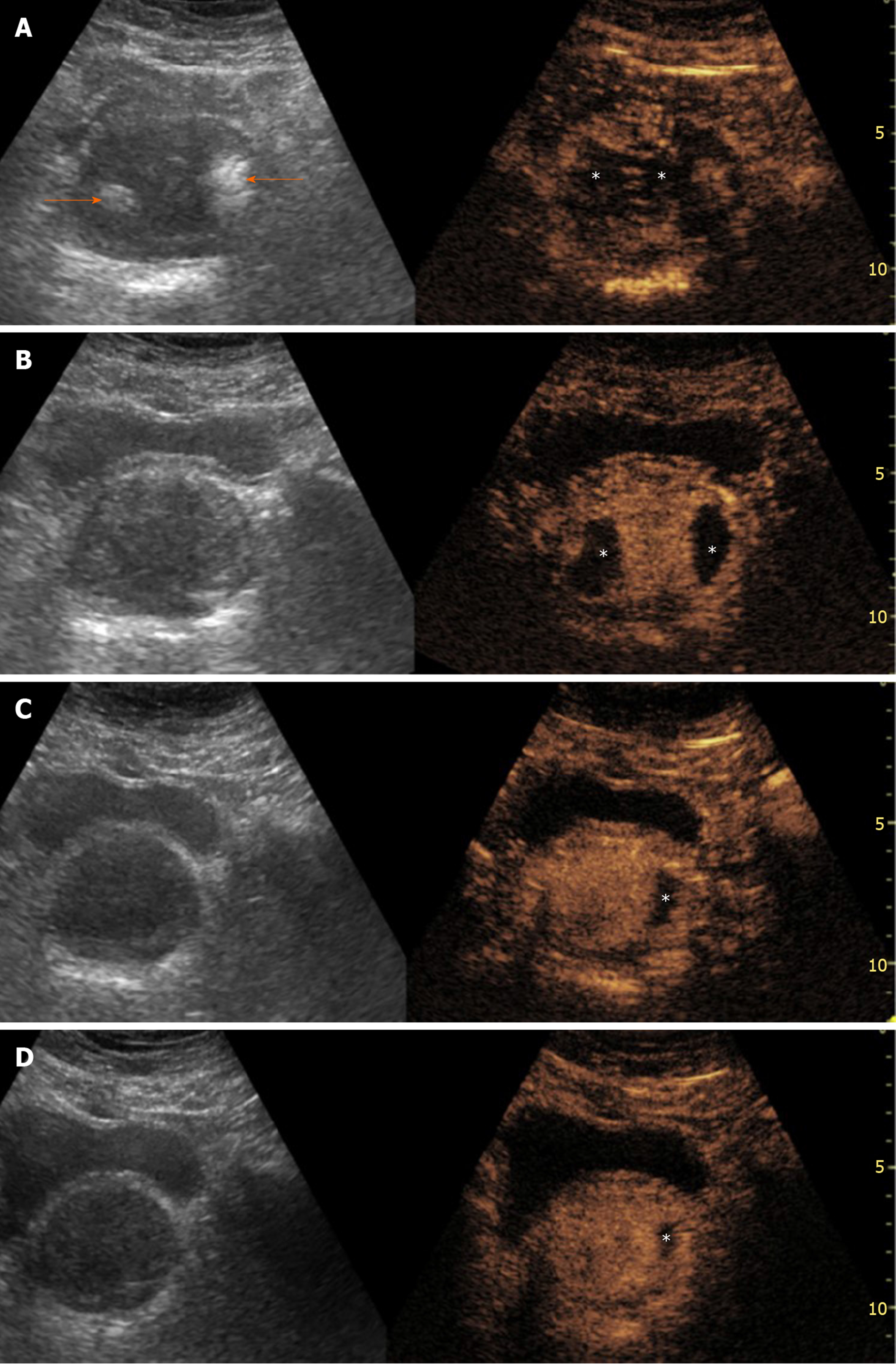
Figure 1 Evolution of ultrasonography and contrast-enhanced ultrasonography findings post-prostatic artery embolization.
Findings are presented in a “split-screen” mode with unenhanced, reference B-mode image on the left, and corresponding contrast-enhanced ultrasonography (CEUS) image on the right. A: One day post-prostatic artery embolization (PAE), transabdominal US (t.a.-US) shows newly appearing echogenic foci, in both prostatic lobes (arrows). t.a.-CEUS shows extensive bilateral prostatic infarction; B: One month post-PAE, the echogenic foci have disappeared and prostatic infarcts are better delineated and smaller in size; C and D: Evaluation 6 and 12 mo post-PAE, respectively, shows that infarcts are diminished or have disappeared and prostate shrinkage can be appreciated. Asterisks indicate prostatic infarcts in all CEUS images. All images are in axial plane.
- Citation: Moschouris H, Dimakis A, Anagnostopoulou A, Stamatiou K, Malagari K. Sonographic evaluation of prostatic artery embolization: Far beyond size measurements. World J Radiol 2020; 12(8): 172-183
- URL: https://www.wjgnet.com/1949-8470/full/v12/i8/172.htm
- DOI: https://dx.doi.org/10.4329/wjr.v12.i8.172


