Copyright
©The Author(s) 2020.
World J Radiol. Aug 28, 2020; 12(8): 156-171
Published online Aug 28, 2020. doi: 10.4329/wjr.v12.i8.156
Published online Aug 28, 2020. doi: 10.4329/wjr.v12.i8.156
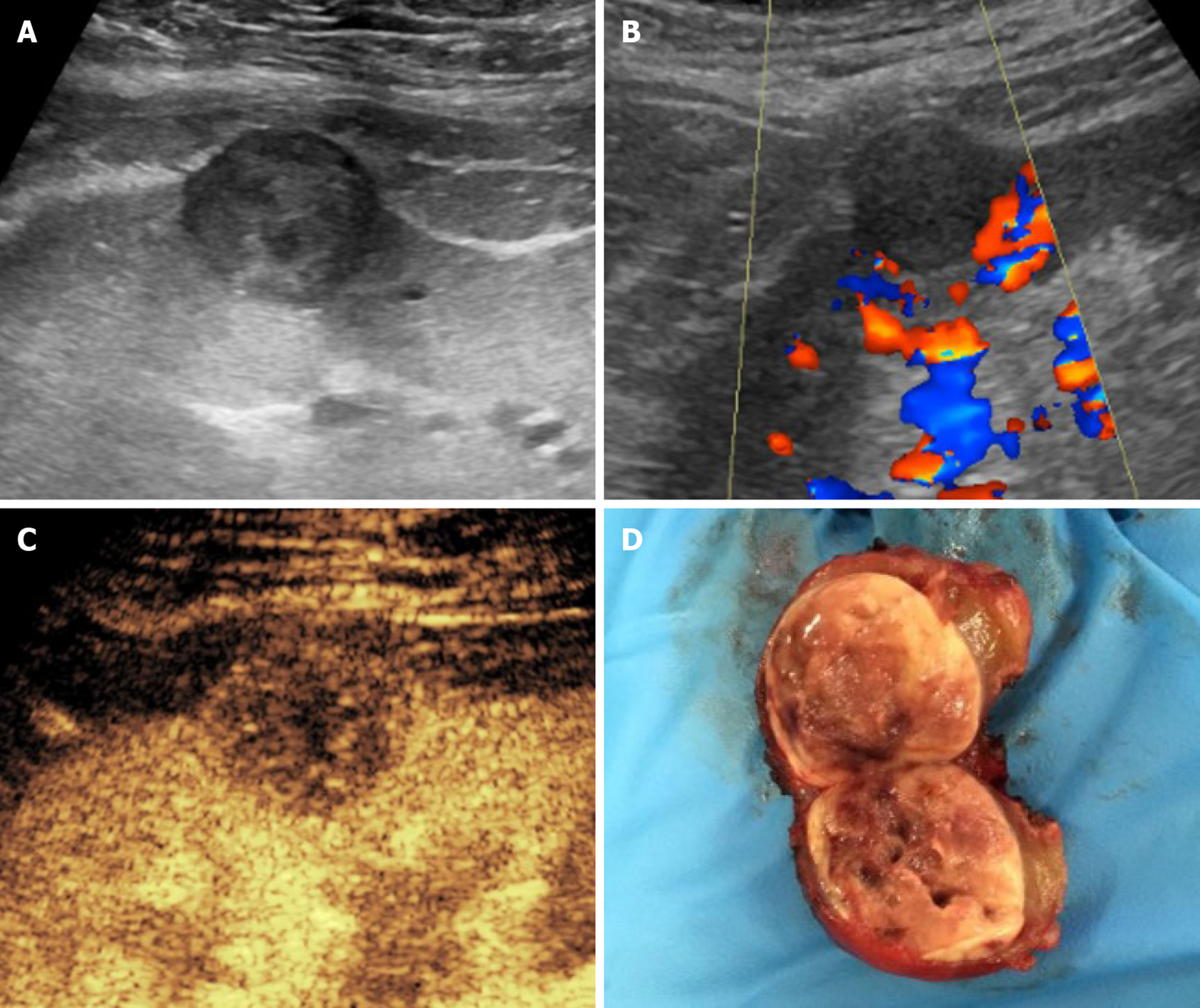
Figure 8 Histologically-proven papillary carcinoma in a 57-year-old male patient with previous kidney transplantation and impaired renal function.
A and B: On grayscale ultrasound (A) the lesion appeared as a hypoechoic cortical mass with absent vascular signal at color analysis (B); C: Contrast-enhanced ultrasound better assessed the solid nature of the mass by showing lower lesion vascularization as compared to normal graft parenchyma; D: The patient was referred to surgery without the need of performing computed tomography or magnetic resonance imaging.
- Citation: Como G, Da Re J, Adani GL, Zuiani C, Girometti R. Role for contrast-enhanced ultrasound in assessing complications after kidney transplant. World J Radiol 2020; 12(8): 156-171
- URL: https://www.wjgnet.com/1949-8470/full/v12/i8/156.htm
- DOI: https://dx.doi.org/10.4329/wjr.v12.i8.156


