Copyright
©The Author(s) 2020.
World J Radiol. Aug 28, 2020; 12(8): 156-171
Published online Aug 28, 2020. doi: 10.4329/wjr.v12.i8.156
Published online Aug 28, 2020. doi: 10.4329/wjr.v12.i8.156
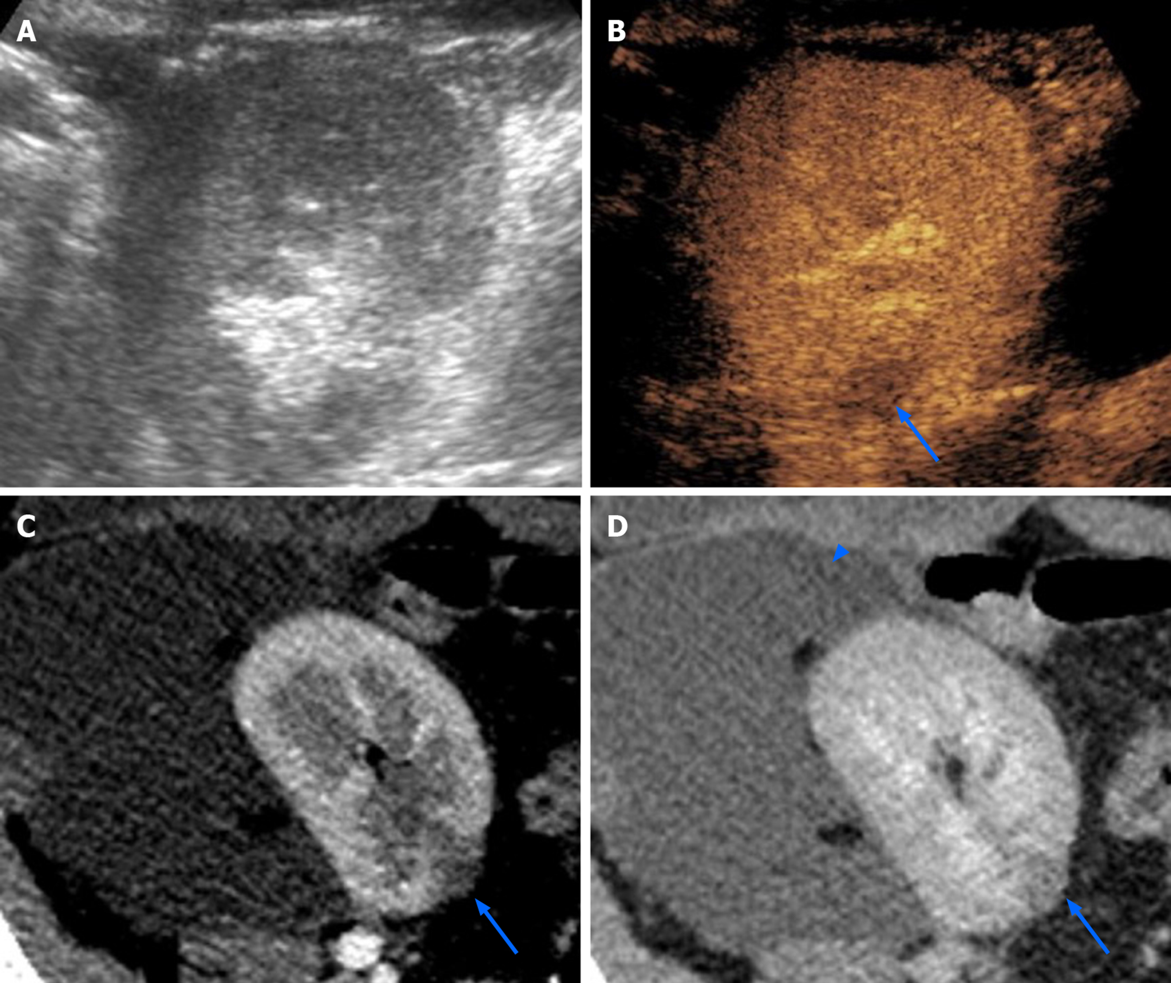
Figure 7 Focal pyelonephritis occurring 50 d after kidney transplantation in a 34-year-old female patient.
While grayscale ultrasound found no abnormalities (A), contrast-enhanced ultrasound revealed a focal hypoperfused area (arrow in B) that was subsequently confirmed on both corticomedullary phase (arrow in C) and nephrographic phase (arrow in D) of a computed tomography performed to assess a large symptomatic perirenal collection (arrowhead in D).
- Citation: Como G, Da Re J, Adani GL, Zuiani C, Girometti R. Role for contrast-enhanced ultrasound in assessing complications after kidney transplant. World J Radiol 2020; 12(8): 156-171
- URL: https://www.wjgnet.com/1949-8470/full/v12/i8/156.htm
- DOI: https://dx.doi.org/10.4329/wjr.v12.i8.156


