Copyright
©The Author(s) 2020.
World J Radiol. Aug 28, 2020; 12(8): 156-171
Published online Aug 28, 2020. doi: 10.4329/wjr.v12.i8.156
Published online Aug 28, 2020. doi: 10.4329/wjr.v12.i8.156
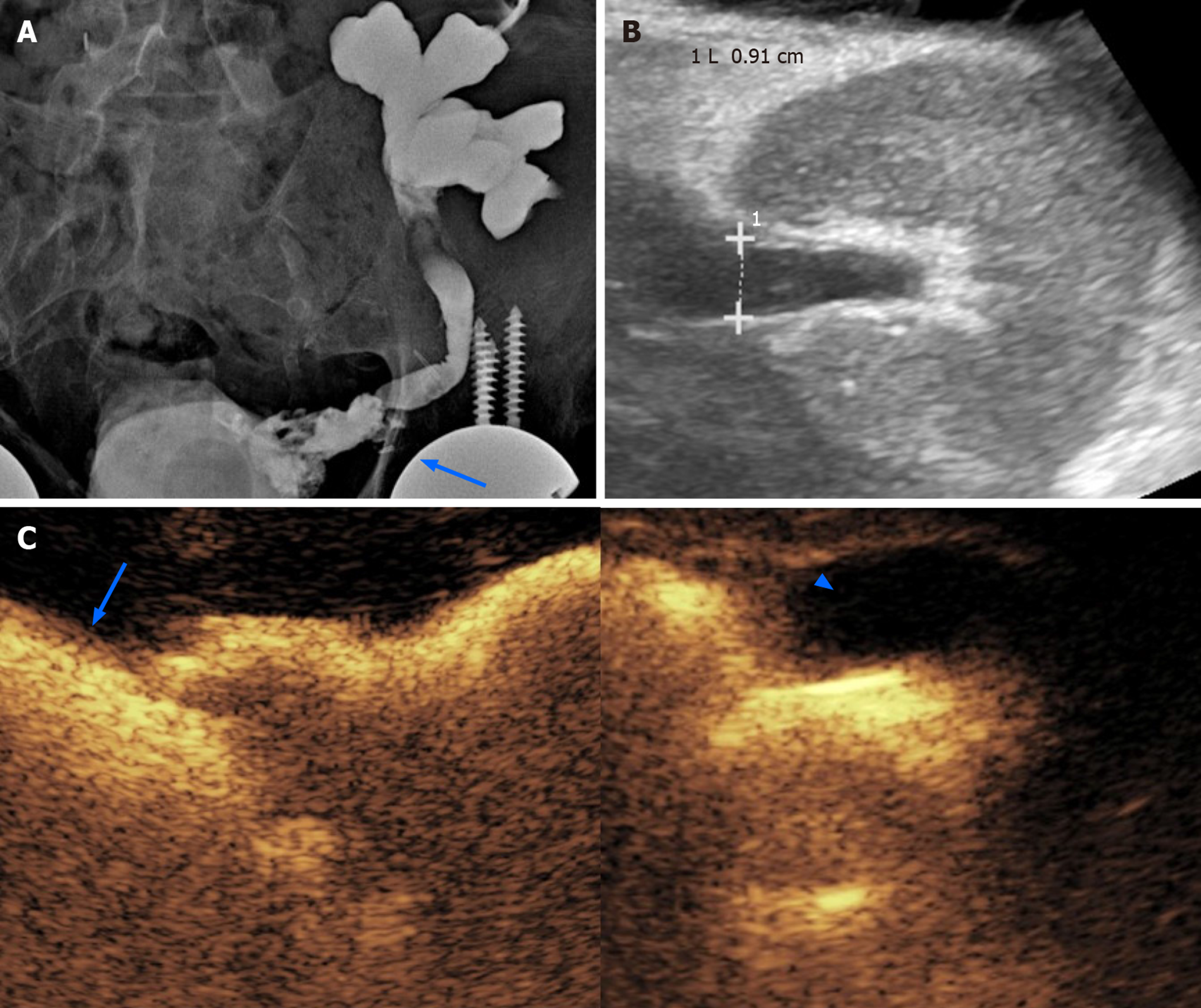
Figure 5 Obstructive renal failure occurring 3 mo after the transplantation in a 62-year-old male patient.
A: A preliminary X-ray nephrostogram showed hydronephrosis with a suspicious urinary leakage at the vesico-ureteral anastomosis (arrow); B: After a conservative management of a few days, the patient was revaluated with grayscale ultrasound, showing reduced hydronephrosis; C: Subsequent contrast-enhanced ultrasound (CEUS)-nephrostogram allowed a panoramic representation of the non-dilated excretory system from the graft (arrowhead) to the bladder (arrow). Based on the resolution of dilation found on CEUS, nephrostomy was successfully removed.
- Citation: Como G, Da Re J, Adani GL, Zuiani C, Girometti R. Role for contrast-enhanced ultrasound in assessing complications after kidney transplant. World J Radiol 2020; 12(8): 156-171
- URL: https://www.wjgnet.com/1949-8470/full/v12/i8/156.htm
- DOI: https://dx.doi.org/10.4329/wjr.v12.i8.156


