Copyright
©The Author(s) 2020.
World J Radiol. Aug 28, 2020; 12(8): 156-171
Published online Aug 28, 2020. doi: 10.4329/wjr.v12.i8.156
Published online Aug 28, 2020. doi: 10.4329/wjr.v12.i8.156
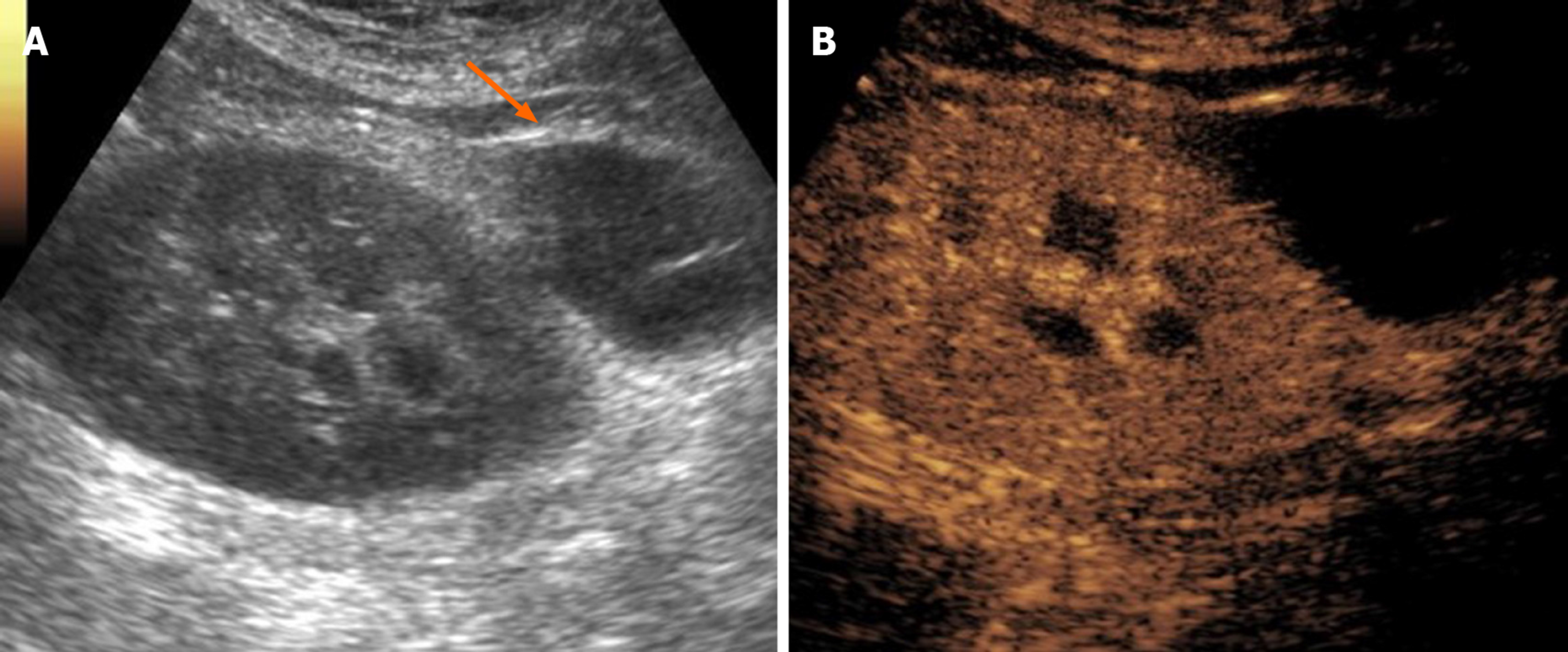
Figure 4 Postoperative lymphocele in a 62-year-old patient who underwent cadaveric kidney transplantation for end-stage renal disease.
A: On B-mode ultrasound, lymphocele was visible as an anechoic collection in the perirenal space (arrow), close to the external iliac vessels of the donor; B: Contrast injection reinforced diagnosis by showing absent vascularization and improved conspicuity with better delineation of the collection size and anatomical relationships.
- Citation: Como G, Da Re J, Adani GL, Zuiani C, Girometti R. Role for contrast-enhanced ultrasound in assessing complications after kidney transplant. World J Radiol 2020; 12(8): 156-171
- URL: https://www.wjgnet.com/1949-8470/full/v12/i8/156.htm
- DOI: https://dx.doi.org/10.4329/wjr.v12.i8.156


