Copyright
©The Author(s) 2020.
World J Radiol. Aug 28, 2020; 12(8): 156-171
Published online Aug 28, 2020. doi: 10.4329/wjr.v12.i8.156
Published online Aug 28, 2020. doi: 10.4329/wjr.v12.i8.156
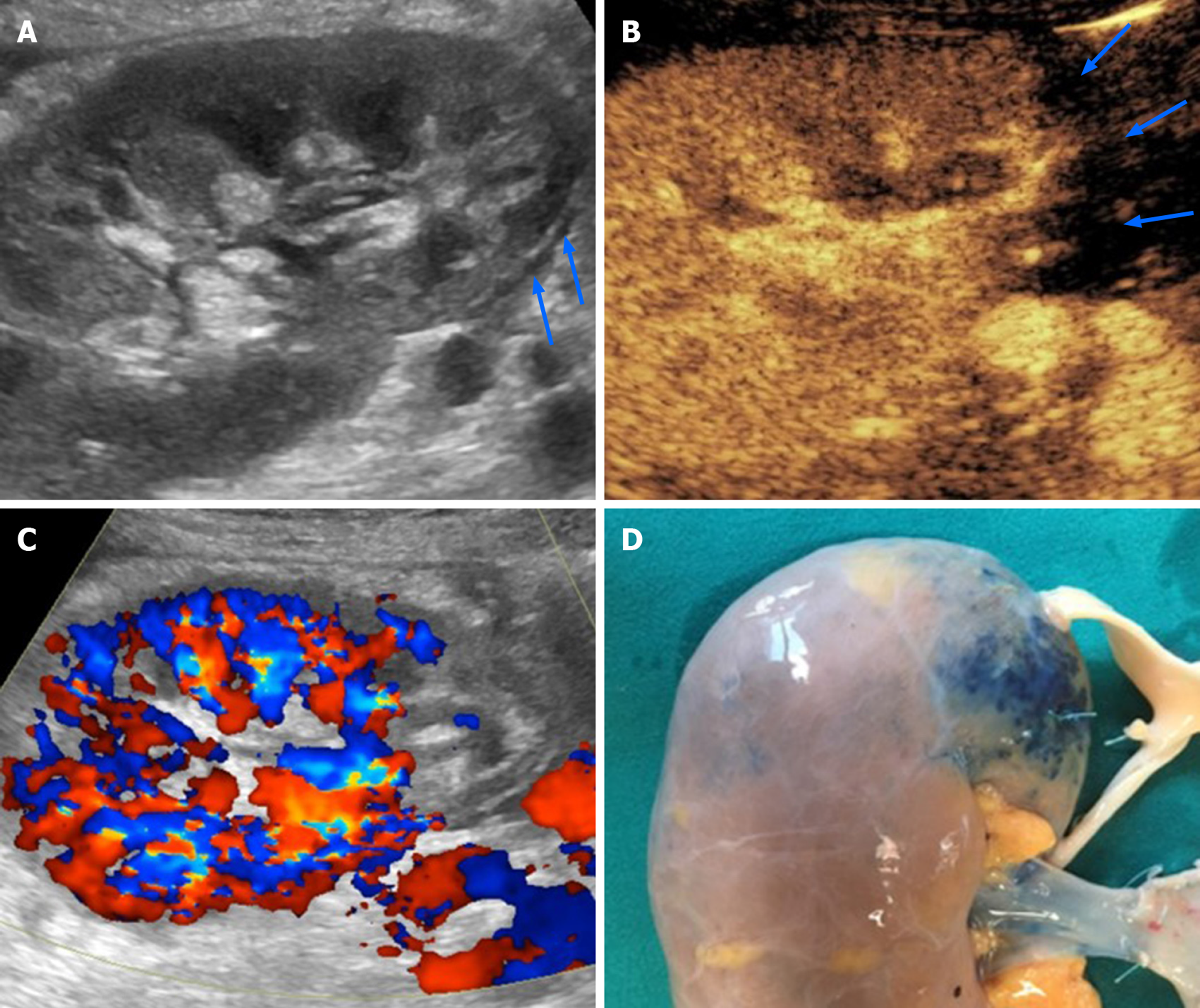
Figure 3 Renal infarction occurring 4 d after cadaveric kidney transplantation in a 53-year-old patient with early renal graft dysfunction.
A and B: Grayscale ultrasound found an area of patchy echotexture (arrows in A) showing absent perfusion on contrast-enhanced ultrasound pulse inversion mode (arrows in B); C: Of note, contrast administration enhanced color signal, thus making this area clearly visible even with this technique; D: Infarction reasonably occurred in the blue-marked area in the pre-transplant image of the graft. This area was served by a small polar artery for which anastomosis was impossible.
- Citation: Como G, Da Re J, Adani GL, Zuiani C, Girometti R. Role for contrast-enhanced ultrasound in assessing complications after kidney transplant. World J Radiol 2020; 12(8): 156-171
- URL: https://www.wjgnet.com/1949-8470/full/v12/i8/156.htm
- DOI: https://dx.doi.org/10.4329/wjr.v12.i8.156


