Copyright
©The Author(s) 2020.
World J Radiol. Jun 28, 2020; 12(6): 87-100
Published online Jun 28, 2020. doi: 10.4329/wjr.v12.i6.87
Published online Jun 28, 2020. doi: 10.4329/wjr.v12.i6.87
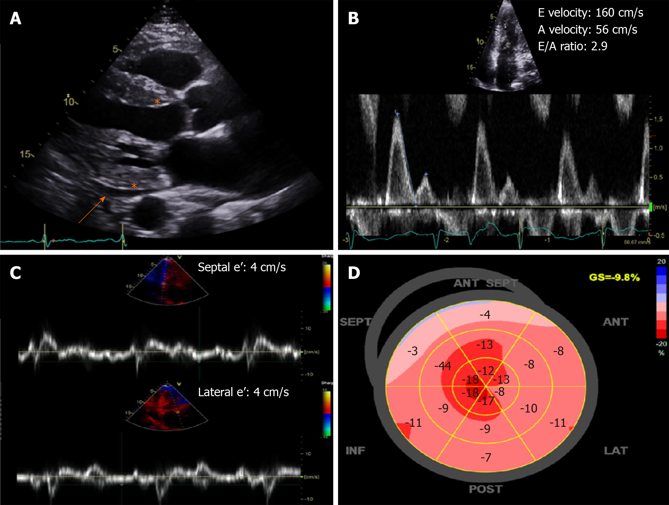
Figure 2 Characteristic echocardiography findings of cardiac amyloidosis.
A: Increased left ventricular wall thickness and echogenicity (asterisk) with small effusion (orange arrow); B and C: Severe (restrictive) diastolic dysfunction with mitral inflow E/A ratio > 2.0 and low septal and lateral e’ velocities by tissue Doppler; D: Bull’s eye plot showing a “cherry-top” pattern of left ventricular peak systolic longitudinal strain, consistent with apical sparing pattern.
- Citation: Wang TKM, Abou Hassan OK, Jaber W, Xu B. Multi-modality imaging of cardiac amyloidosis: Contemporary update. World J Radiol 2020; 12(6): 87-100
- URL: https://www.wjgnet.com/1949-8470/full/v12/i6/87.htm
- DOI: https://dx.doi.org/10.4329/wjr.v12.i6.87


