Copyright
©The Author(s) 2020.
World J Radiol. May 28, 2020; 12(5): 76-86
Published online May 28, 2020. doi: 10.4329/wjr.v12.i5.76
Published online May 28, 2020. doi: 10.4329/wjr.v12.i5.76
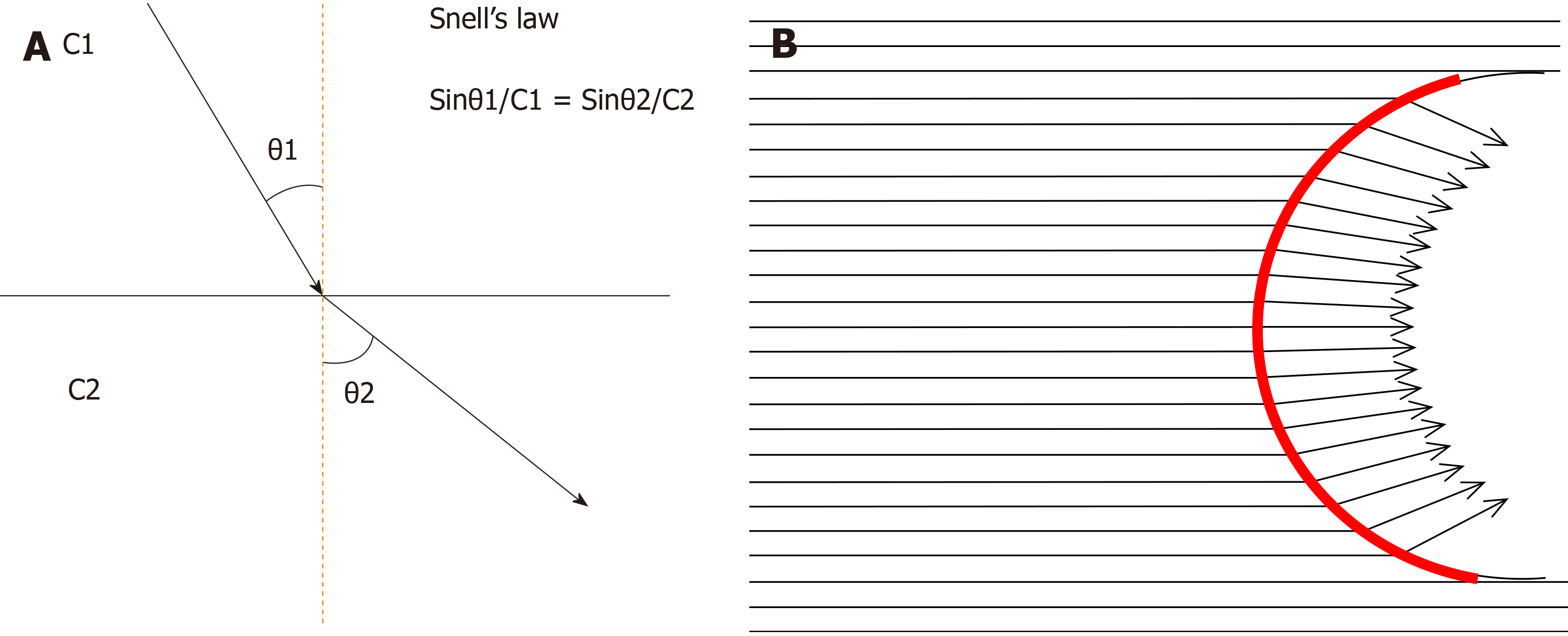
Figure 10 Sound refraction at the interface.
A: Sound (or shear wave) refraction occurs at the straight interface between two structures of different acoustic (or shear wave) velocities, depending on Snell’s law. The degree of refraction is always the same. θ1: Angle of incidence, θ2: Angle of refraction, C1: Sound (or shear wave) velocity in tissue 1, C2: Sound (or shear wave) velocity in tissue 2; B: Sound (or shear wave) refraction at the curved interface. As the tumor (or cirrhotic liver surface) has a curved interface, the angle of incidence changes according to the incidental point. Thus, the angle of refraction changes also according to the incidental point. Red line: Curved interface.
- Citation: Naganuma H, Ishida H, Uno A, Nagai H, Kuroda H, Ogawa M. Diagnostic problems in two-dimensional shear wave elastography of the liver. World J Radiol 2020; 12(5): 76-86
- URL: https://www.wjgnet.com/1949-8470/full/v12/i5/76.htm
- DOI: https://dx.doi.org/10.4329/wjr.v12.i5.76


