Copyright
©The Author(s) 2020.
World J Radiol. May 28, 2020; 12(5): 76-86
Published online May 28, 2020. doi: 10.4329/wjr.v12.i5.76
Published online May 28, 2020. doi: 10.4329/wjr.v12.i5.76
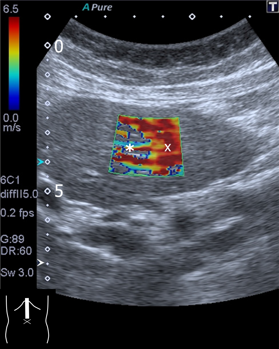
Figure 5 Motion artifact.
Two-dimensional shear wave elastography of the liver left lobe shows this characteristic finding of a colorless area (asterisk), suggesting inaccurate measurement of shear wave values. The reddish part (×) of the cursor is also unreliable.
- Citation: Naganuma H, Ishida H, Uno A, Nagai H, Kuroda H, Ogawa M. Diagnostic problems in two-dimensional shear wave elastography of the liver. World J Radiol 2020; 12(5): 76-86
- URL: https://www.wjgnet.com/1949-8470/full/v12/i5/76.htm
- DOI: https://dx.doi.org/10.4329/wjr.v12.i5.76


