Copyright
©The Author(s) 2020.
World J Radiol. May 28, 2020; 12(5): 48-67
Published online May 28, 2020. doi: 10.4329/wjr.v12.i5.48
Published online May 28, 2020. doi: 10.4329/wjr.v12.i5.48
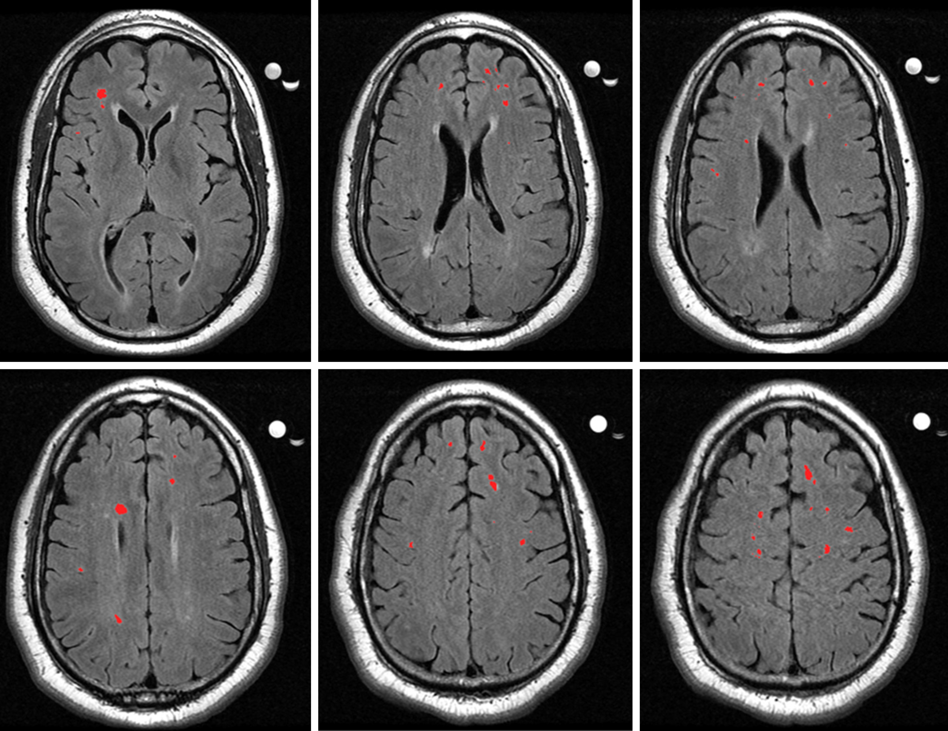
Figure 3 Axial slices from a magnetic resonance imaging scan of a vascular depression patient.
The highlighted sections are deep white matter hyperintensities, which have been graded as a severity of 2 on the Fazekas rating scale.
- Citation: Rushia SN, Shehab AAS, Motter JN, Egglefield DA, Schiff S, Sneed JR, Garcon E. Vascular depression for radiology: A review of the construct, methodology, and diagnosis. World J Radiol 2020; 12(5): 48-67
- URL: https://www.wjgnet.com/1949-8470/full/v12/i5/48.htm
- DOI: https://dx.doi.org/10.4329/wjr.v12.i5.48


