Copyright
©The Author(s) 2020.
World J Radiol. Apr 28, 2020; 12(4): 29-47
Published online Apr 28, 2020. doi: 10.4329/wjr.v12.i4.29
Published online Apr 28, 2020. doi: 10.4329/wjr.v12.i4.29
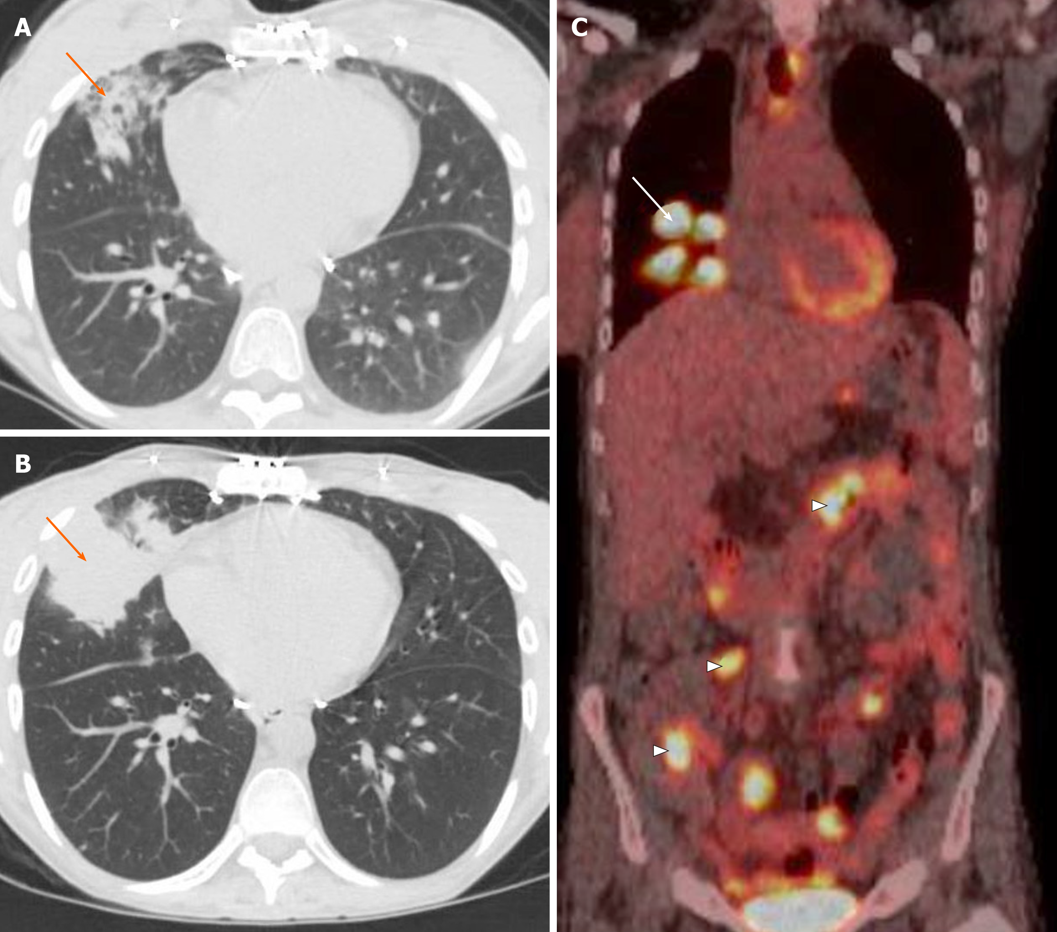
Figure 14 Post-transplant lymphoproliferative disease.
A: A 31-year-old woman with history of cystic fibrosis and double lung transplant presented with cough and computed tomography of the chest demonstrated consolidation in right middle lobe; B: Despite treatment, follow-up computed tomography after 2 mo demonstrated worsening right middle lobe consolidation (arrow); C: Fluorodeoxyglucose-positron emission tomography confirmed intense metabolic activity in the lung opacity (arrow) as well as areas of increased metabolic activity in the bowel (arrowheads). Biopsy confirmed the post-transplant lymphoproliferative disease.
- Citation: Ansari-Gilani K, Chalian H, Rassouli N, Bedayat A, Kalisz K. Chronic airspace disease: Review of the causes and key computed tomography findings. World J Radiol 2020; 12(4): 29-47
- URL: https://www.wjgnet.com/1949-8470/full/v12/i4/29.htm
- DOI: https://dx.doi.org/10.4329/wjr.v12.i4.29


