Copyright
©The Author(s) 2020.
World J Radiol. Apr 28, 2020; 12(4): 29-47
Published online Apr 28, 2020. doi: 10.4329/wjr.v12.i4.29
Published online Apr 28, 2020. doi: 10.4329/wjr.v12.i4.29
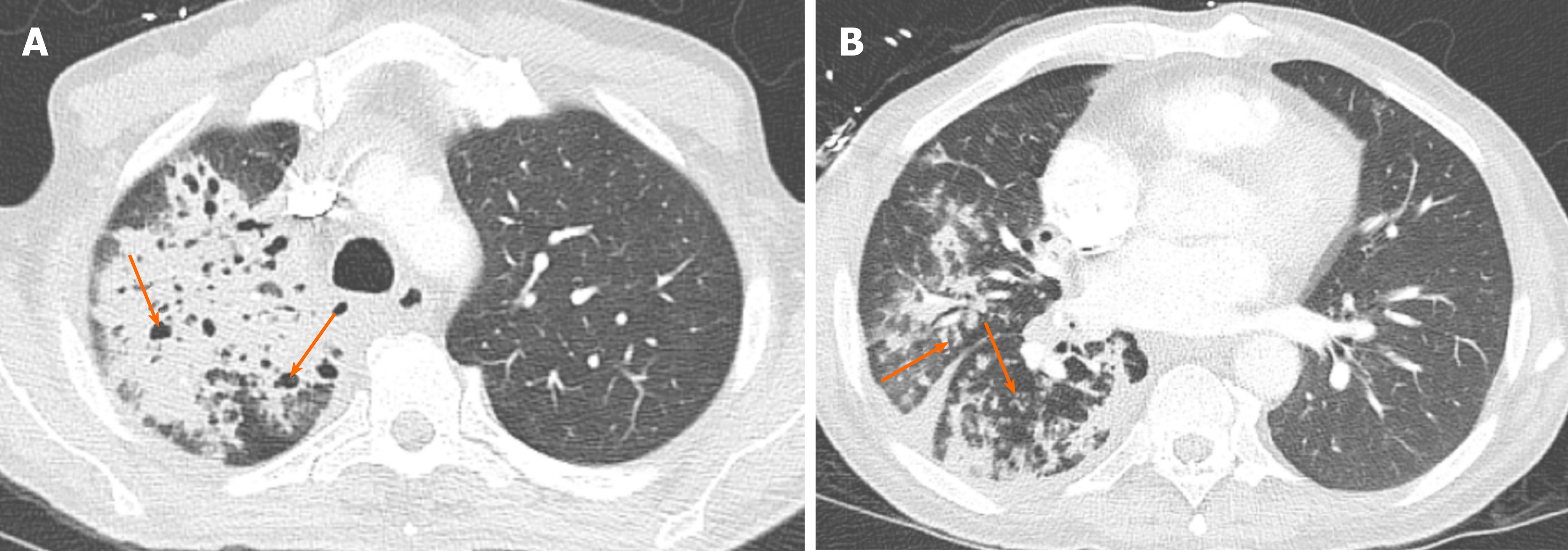
Figure 8 Mycobacterial tuberculosis.
A 49-year-old man with cough and hemoptysis. A: Large area of consolidation was present in the right upper lobe, with small areas of cavitation (arrows); B: There was a significant amount of airspace opacity in the ipsilateral lung involving all three lobes, with areas of tree-in-bud nodularity (arrows) keeping with an endobronchial spread of infection.
- Citation: Ansari-Gilani K, Chalian H, Rassouli N, Bedayat A, Kalisz K. Chronic airspace disease: Review of the causes and key computed tomography findings. World J Radiol 2020; 12(4): 29-47
- URL: https://www.wjgnet.com/1949-8470/full/v12/i4/29.htm
- DOI: https://dx.doi.org/10.4329/wjr.v12.i4.29


