Copyright
©The Author(s) 2020.
World J Radiol. Apr 28, 2020; 12(4): 29-47
Published online Apr 28, 2020. doi: 10.4329/wjr.v12.i4.29
Published online Apr 28, 2020. doi: 10.4329/wjr.v12.i4.29
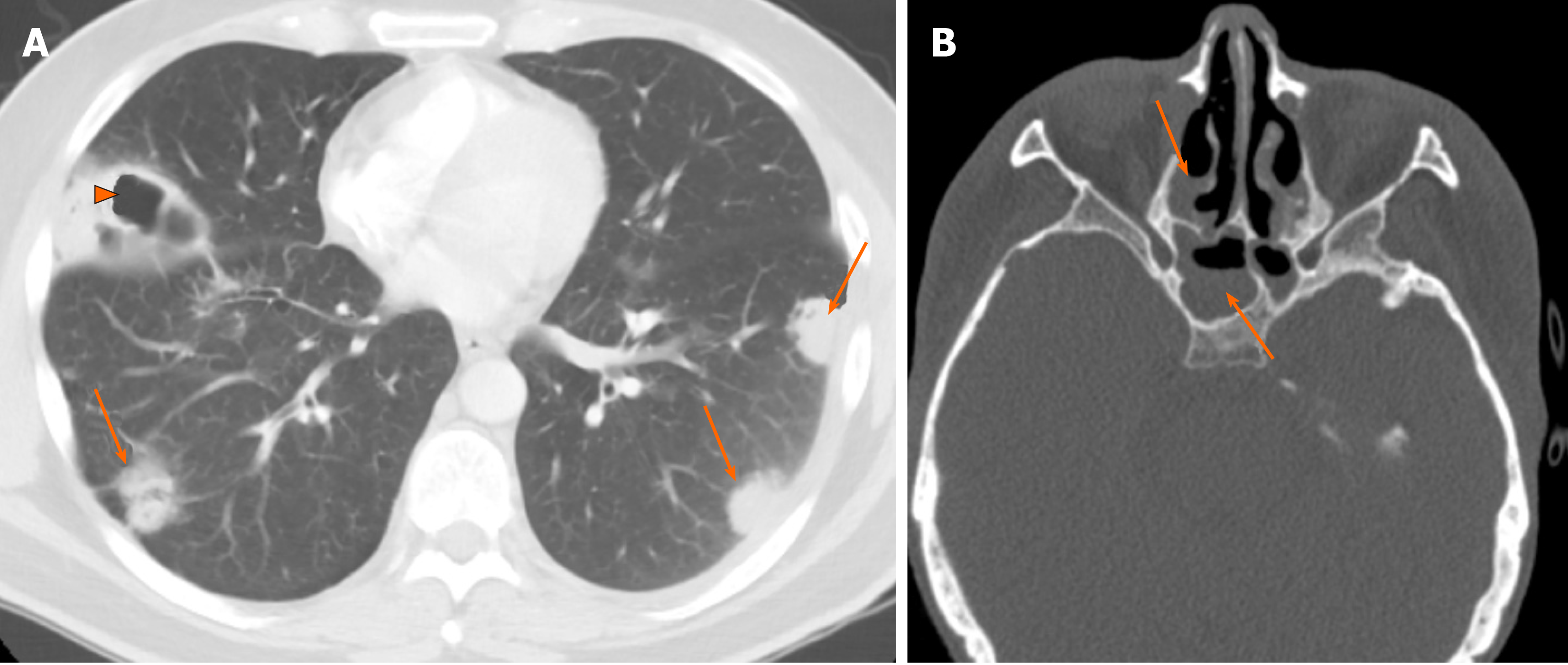
Figure 5 Granulomatosis with polyangiitis.
A 38-year-old male with hemoptysis and shortness of breath. A: Non-contrast enhanced computed tomography of the chest (A) showed multiple randomly distributed nodules/masses and round consolidative opacities in both lungs (arrows), with areas of cavitation (arrowheads); B: Head computed tomography showed involvement of bilateral sphenoid sinuses and ethmoidal air cells (arrows) in keeping with pan-sinusitis.
- Citation: Ansari-Gilani K, Chalian H, Rassouli N, Bedayat A, Kalisz K. Chronic airspace disease: Review of the causes and key computed tomography findings. World J Radiol 2020; 12(4): 29-47
- URL: https://www.wjgnet.com/1949-8470/full/v12/i4/29.htm
- DOI: https://dx.doi.org/10.4329/wjr.v12.i4.29


