Copyright
©The Author(s) 2020.
World J Radiol. Apr 28, 2020; 12(4): 29-47
Published online Apr 28, 2020. doi: 10.4329/wjr.v12.i4.29
Published online Apr 28, 2020. doi: 10.4329/wjr.v12.i4.29
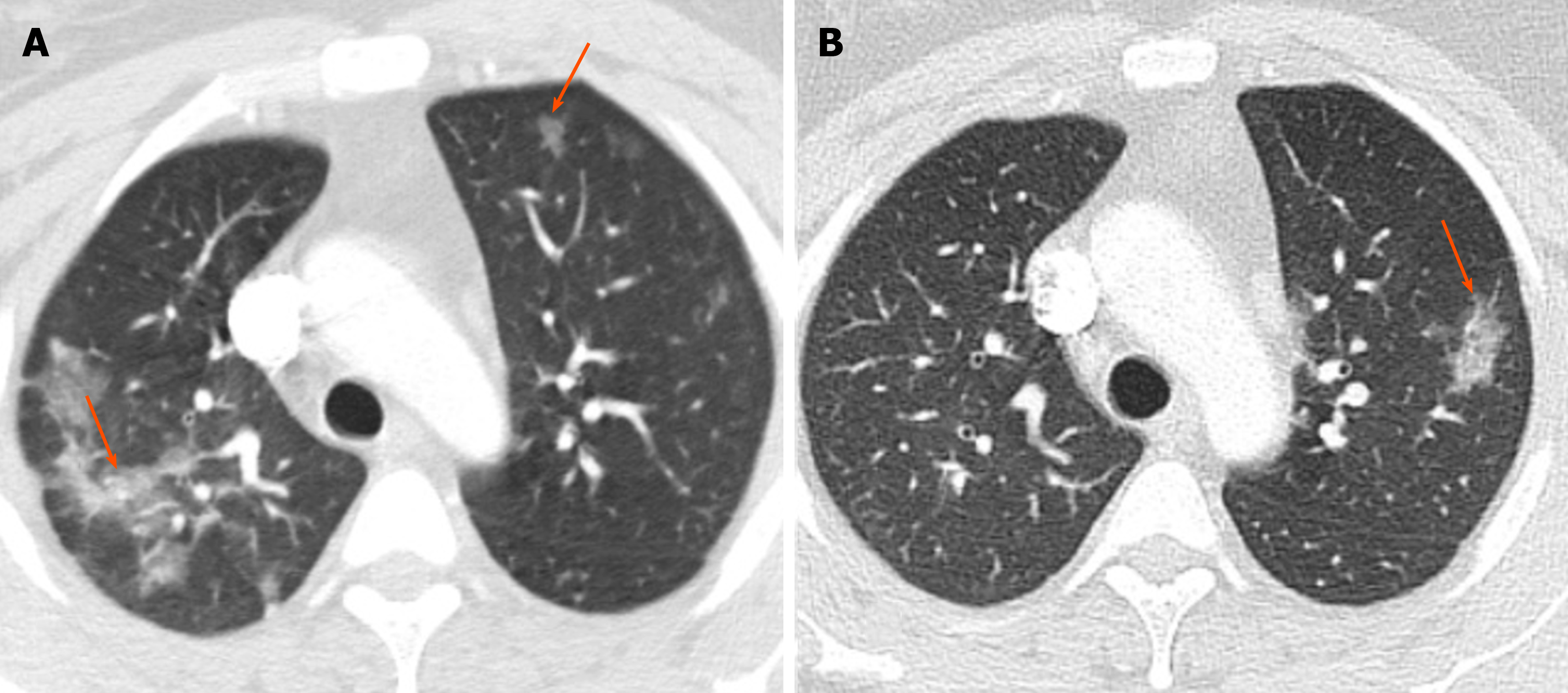
Figure 2 Chronic eosinophilic pneumonia.
A: Baseline non-contrast chest computed tomography in a 38-year-old woman with history of asthma, eosinophilia and eosinophilic pneumonia, who presented with shortness of breath, showed bilateral patchy airspace opacities in both upper lobes (arrows); B: Follow-up chest computed tomography after 8 mo showed resolution of the previous opacities with interval development of an area of patchy consolidation in the left upper lobe (arrow).
- Citation: Ansari-Gilani K, Chalian H, Rassouli N, Bedayat A, Kalisz K. Chronic airspace disease: Review of the causes and key computed tomography findings. World J Radiol 2020; 12(4): 29-47
- URL: https://www.wjgnet.com/1949-8470/full/v12/i4/29.htm
- DOI: https://dx.doi.org/10.4329/wjr.v12.i4.29


