Copyright
©The Author(s) 2020.
World J Radiology. Mar 28, 2020; 12(3): 18-28
Published online Mar 28, 2020. doi: 10.4329/wjr.v12.i3.18
Published online Mar 28, 2020. doi: 10.4329/wjr.v12.i3.18
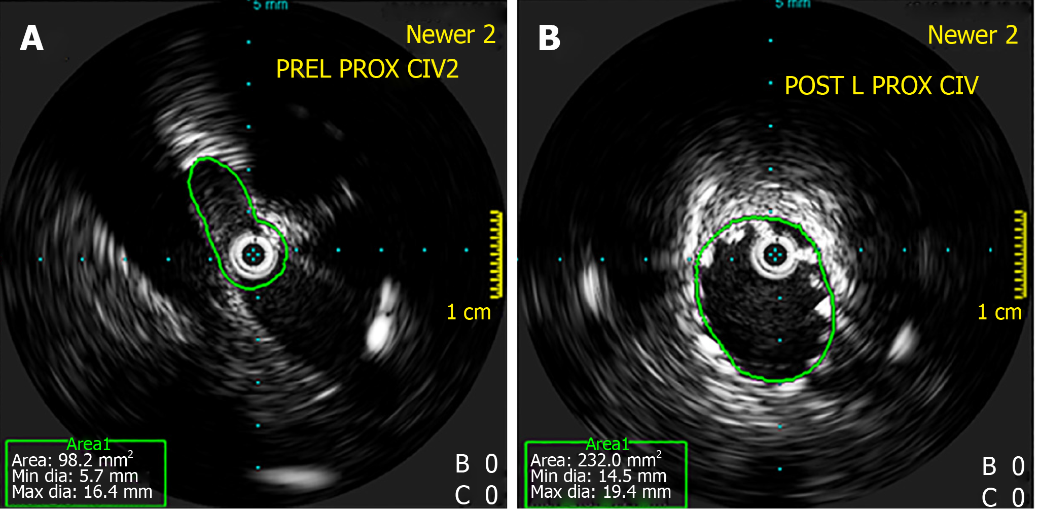
Figure 5 Intravascular ultrasound.
A: Intravascular ultrasound showing stenosis at the left proximal common iliac vein; B: Intravascular ultrasound showing dilatation post-stenting. Intravascular ultrasound allows for direct imaging of the stenosis and evaluation of post-stenting vein diameter.
- Citation: Toh MR, Tang TY, Lim HHMN, Venkatanarasimha N, Damodharan K. Review of imaging and endovascular intervention of iliocaval venous compression syndrome. World J Radiology 2020; 12(3): 18-28
- URL: https://www.wjgnet.com/1949-8470/full/v12/i3/18.htm
- DOI: https://dx.doi.org/10.4329/wjr.v12.i3.18


