Copyright
©The Author(s) 2020.
World J Radiology. Mar 28, 2020; 12(3): 18-28
Published online Mar 28, 2020. doi: 10.4329/wjr.v12.i3.18
Published online Mar 28, 2020. doi: 10.4329/wjr.v12.i3.18
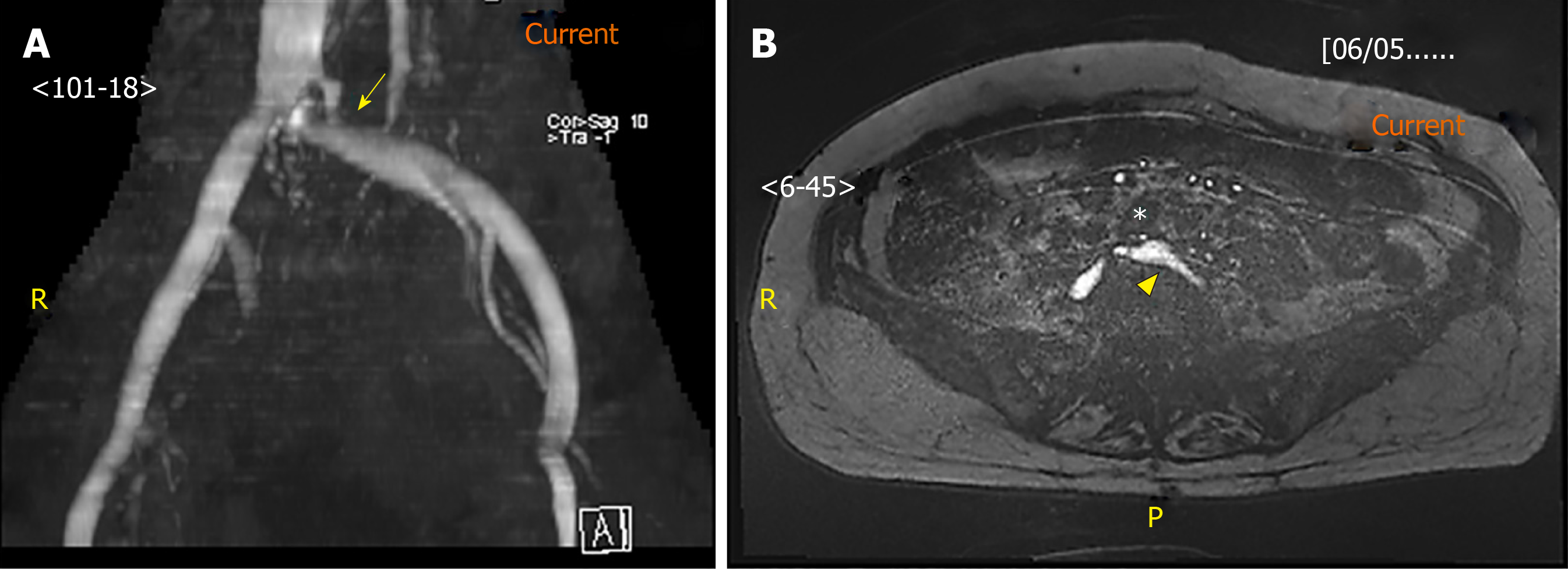
Figure 2 Magnetic resonance venography image.
A: Magnetic resonance venography image of the stenosed left common iliac vein (arrow); B: Axial image of the left common iliac artery (asterisk) compressing against the left common iliac vein (arrowhead). Under magnetic resonance venography, venous blood generates high signal intensity (hyperintense) while arterial blood is suppressed (hypointense).
- Citation: Toh MR, Tang TY, Lim HHMN, Venkatanarasimha N, Damodharan K. Review of imaging and endovascular intervention of iliocaval venous compression syndrome. World J Radiology 2020; 12(3): 18-28
- URL: https://www.wjgnet.com/1949-8470/full/v12/i3/18.htm
- DOI: https://dx.doi.org/10.4329/wjr.v12.i3.18


