Copyright
©The Author(s) 2020.
World J Radiol. Feb 28, 2020; 12(2): 10-17
Published online Feb 28, 2020. doi: 10.4329/wjr.v12.i2.10
Published online Feb 28, 2020. doi: 10.4329/wjr.v12.i2.10
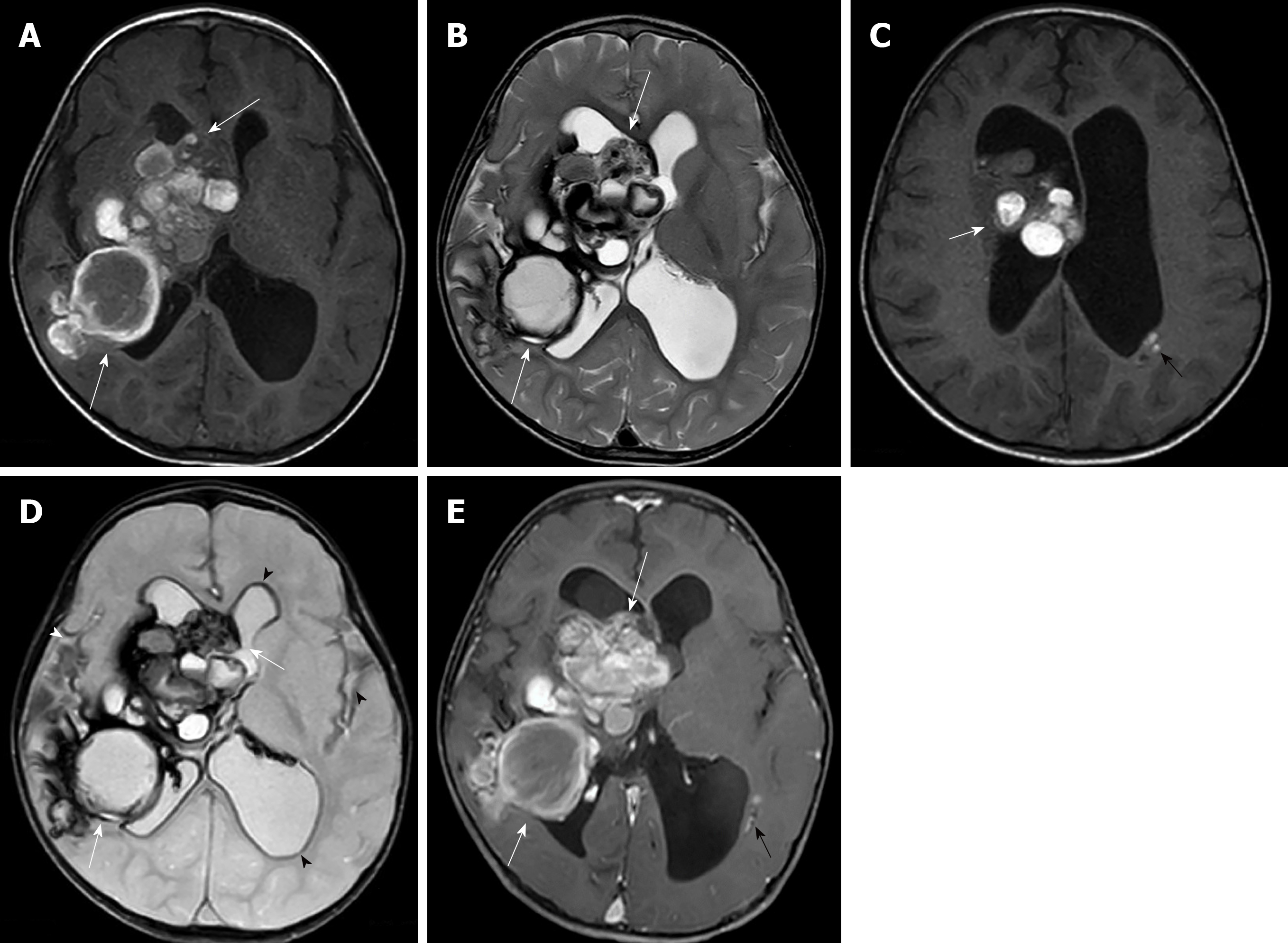
Figure 3 Pre-operative magnetic resonance imaging.
A: Axial T1 weighted image; B: Axial T2 weighted image; C: Axial FLAIR image; D: Axial T2* weighted image; E: Axial T1 weighted post-contrast image show multifocal multilobulated cystic masses with various stages of hemorrhage or bubbles of blood appearance, at the right periventricular region, right foramen of Monro, right trigone, occipital horn of the right lateral ventricle (white arrows) and body of left lateral ventricle (black arrow) with moderate hydrocephalus. Old intraventricular hemorrhage and superficial siderosis (arrowheads) at both fronto-temporal sulci and all subependymal lining are in axial T2* weighted image.
- Citation: Eng-Chuan S, Kritsaneepaiboon S, Kaewborisutsakul A, Kanjanapradit K. Giant intraventricular and paraventricular cavernous malformations with multifocal subependymal cavernous malformations in pediatric patients: Two case reports. World J Radiol 2020; 12(2): 10-17
- URL: https://www.wjgnet.com/1949-8470/full/v12/i2/10.htm
- DOI: https://dx.doi.org/10.4329/wjr.v12.i2.10


