Copyright
©The Author(s) 2020.
World J Radiol. Feb 28, 2020; 12(2): 10-17
Published online Feb 28, 2020. doi: 10.4329/wjr.v12.i2.10
Published online Feb 28, 2020. doi: 10.4329/wjr.v12.i2.10
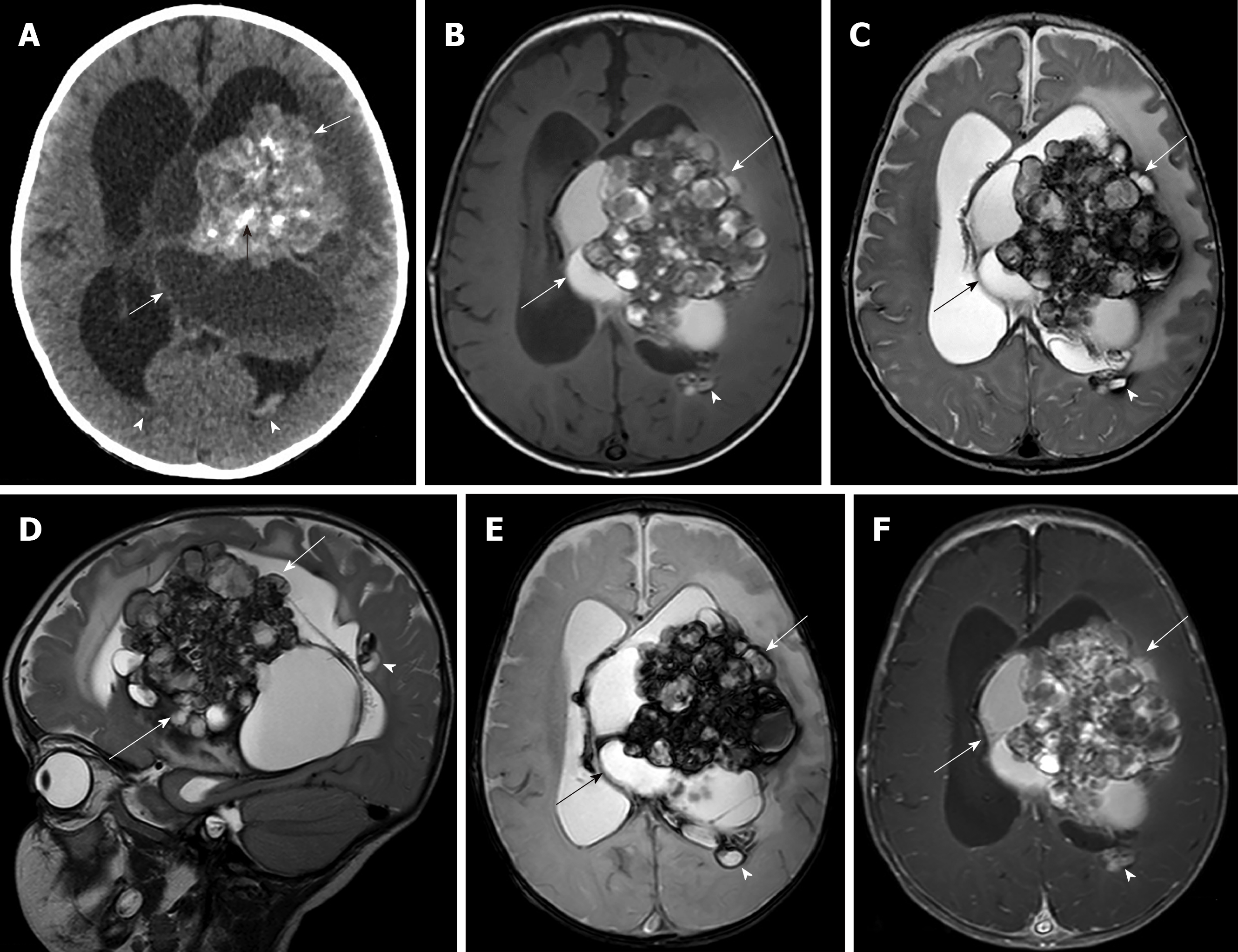
Figure 1 Pre-operative brain computed tomography and magnetic resonance imaging.
A: Non-contrast computed tomography shows cystic-solid mass (white arrows) with internal calcifications (black arrows) in body of left lateral ventricle and intraventricular hemorrhage (arrowheads) resulting in hydrocephalus; B: Axial T1 weighted image; C: Axial T2 weighted image; D: Sagittal T2 weighted image; E: Axial T2* weighted mage; F: Axial T1 weighted post gadolinium administration image show a solid-cystic intraventricular mass (arrows) at the left trigone and body of left lateral ventricle with internal calcification, perilesional brain edema and various stages of hemorrhage; bubbles of blood appearance. Another small slightly enhanced nodule (arrowhead) at the subependymal region of the posterior body of the left lateral ventricle is seen.
- Citation: Eng-Chuan S, Kritsaneepaiboon S, Kaewborisutsakul A, Kanjanapradit K. Giant intraventricular and paraventricular cavernous malformations with multifocal subependymal cavernous malformations in pediatric patients: Two case reports. World J Radiol 2020; 12(2): 10-17
- URL: https://www.wjgnet.com/1949-8470/full/v12/i2/10.htm
- DOI: https://dx.doi.org/10.4329/wjr.v12.i2.10


