Copyright
©The Author(s) 2020.
World J Radiol. Dec 28, 2020; 12(12): 316-326
Published online Dec 28, 2020. doi: 10.4329/wjr.v12.i12.316
Published online Dec 28, 2020. doi: 10.4329/wjr.v12.i12.316
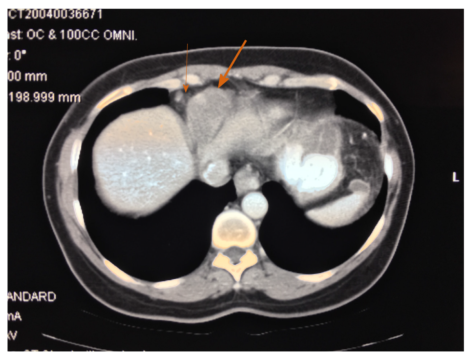
Figure 3 A mass is located directly beneath the diaphragm, invading into the inferior aspect of the pericardial sac and distorting the superior aspect of the liver.
The mass was composed entirely of malignant mesothelioma (large arrow). An enlarged right costophrenic angle lymph node was depicted (small arrow). This disease may be best described as a combined malignant peritoneal and pericardial mesothelioma.
- Citation: Sugarbaker PH, Jelinek JS. Unusual radiologic presentations of malignant peritoneal mesothelioma. World J Radiol 2020; 12(12): 316-326
- URL: https://www.wjgnet.com/1949-8470/full/v12/i12/316.htm
- DOI: https://dx.doi.org/10.4329/wjr.v12.i12.316


