Copyright
©The Author(s) 2020.
World J Radiol. Dec 28, 2020; 12(12): 272-288
Published online Dec 28, 2020. doi: 10.4329/wjr.v12.i12.272
Published online Dec 28, 2020. doi: 10.4329/wjr.v12.i12.272
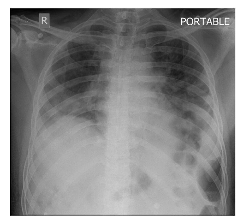
Figure 1 Chest radiograph of 64-year-old coronavirus disease 2019 patient showing peripheral reticular infiltrates and consolidation predominantly in a lower lobe distribution, typical for coronavirus disease 2019 pneumonia.
- Citation: Mathew RP, Jose M, Jayaram V, Joy P, George D, Joseph M, Sleeba T, Toms A. Current status quo on COVID-19 including chest imaging. World J Radiol 2020; 12(12): 272-288
- URL: https://www.wjgnet.com/1949-8470/full/v12/i12/272.htm
- DOI: https://dx.doi.org/10.4329/wjr.v12.i12.272


