Copyright
©The Author(s) 2020.
World J Radiol. Nov 28, 2020; 12(11): 247-260
Published online Nov 28, 2020. doi: 10.4329/wjr.v12.i11.247
Published online Nov 28, 2020. doi: 10.4329/wjr.v12.i11.247
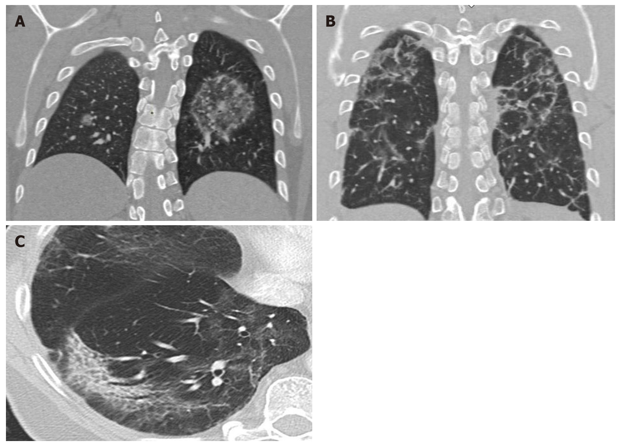
Figure 4 Computed tomography findings late stage disease.
A: Reverse halo or atoll sign: Rounded opacity in the left lung with ground-glass attenuation in the center demarcated by a denser, fine ring. Small, homogeneous rounded opacity in the right lung; B: Bilateral perilobular pattern: Polygonal (irregular, linear or band-like) peripheral opacities in secondary pulmonary lobules; C: Peripheral, elongated, curved consolidation in the right lower lobe containing dilated bronchi.
- Citation: Landete P, Quezada Loaiza CA, Aldave-Orzaiz B, Muñiz SH, Maldonado A, Zamora E, Sam Cerna AC, del Cerro E, Alonso RC, Couñago F. Clinical features and radiological manifestations of COVID-19 disease. World J Radiol 2020; 12(11): 247-260
- URL: https://www.wjgnet.com/1949-8470/full/v12/i11/247.htm
- DOI: https://dx.doi.org/10.4329/wjr.v12.i11.247


