Copyright
©The Author(s) 2020.
World J Radiol. Nov 28, 2020; 12(11): 247-260
Published online Nov 28, 2020. doi: 10.4329/wjr.v12.i11.247
Published online Nov 28, 2020. doi: 10.4329/wjr.v12.i11.247
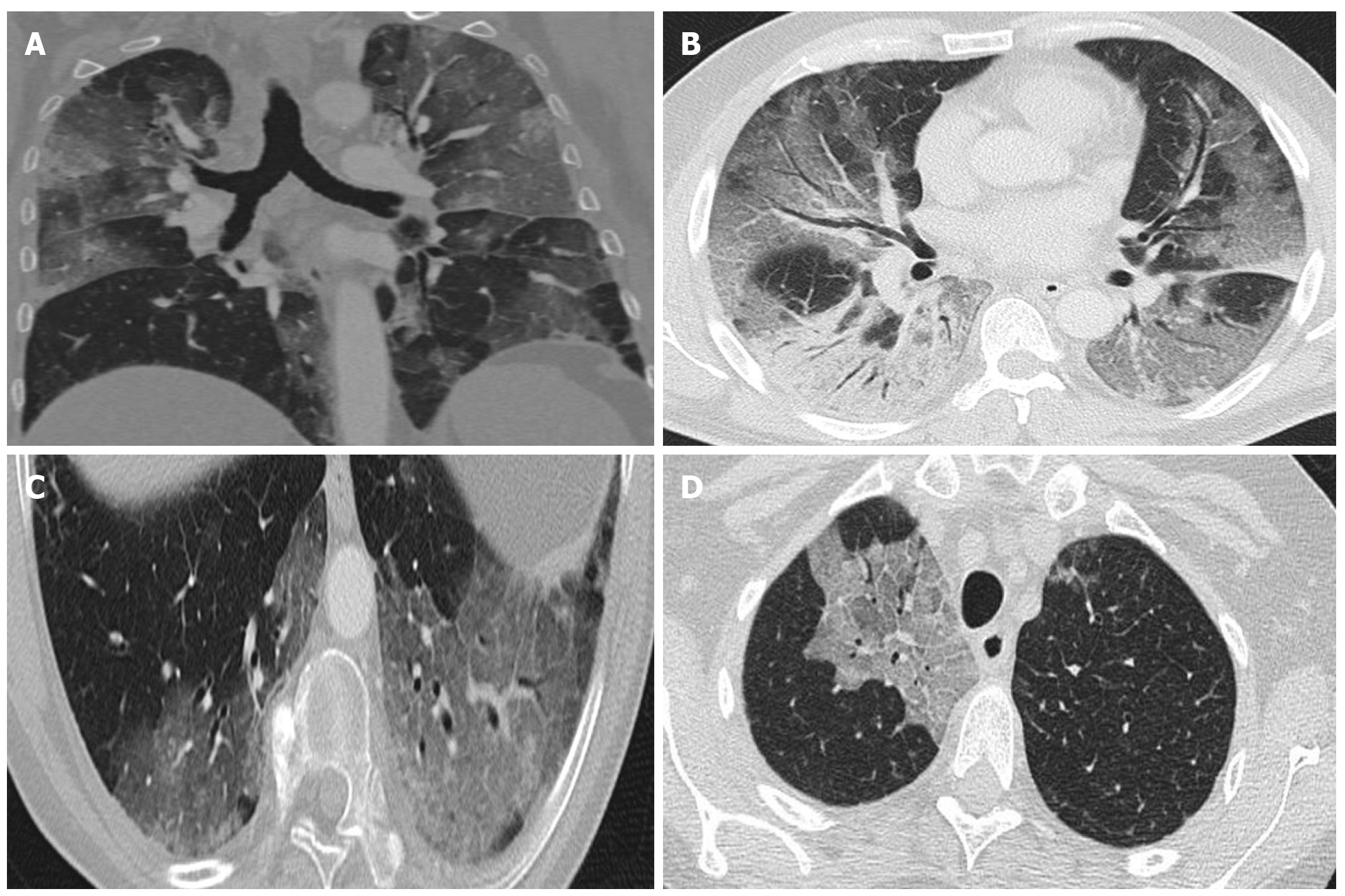
Figure 3 Computed tomography findings.
A: Extensive bilateral ground-glass opacities, one with poorly-defined margins and another with clearly defined borders; B: Multilobar ground-glass opacities and consolidation with air bronchogram in the right lower lobe. Band of subpleural parenchyma respected in the left lung; C: Microvascular dilation sign in the middle of ground-glass opacity in the left lower lobe; D: Crazy-paving pattern in the right upper lobe.
- Citation: Landete P, Quezada Loaiza CA, Aldave-Orzaiz B, Muñiz SH, Maldonado A, Zamora E, Sam Cerna AC, del Cerro E, Alonso RC, Couñago F. Clinical features and radiological manifestations of COVID-19 disease. World J Radiol 2020; 12(11): 247-260
- URL: https://www.wjgnet.com/1949-8470/full/v12/i11/247.htm
- DOI: https://dx.doi.org/10.4329/wjr.v12.i11.247


