Copyright
©The Author(s) 2020.
World J Radiol. Nov 28, 2020; 12(11): 247-260
Published online Nov 28, 2020. doi: 10.4329/wjr.v12.i11.247
Published online Nov 28, 2020. doi: 10.4329/wjr.v12.i11.247
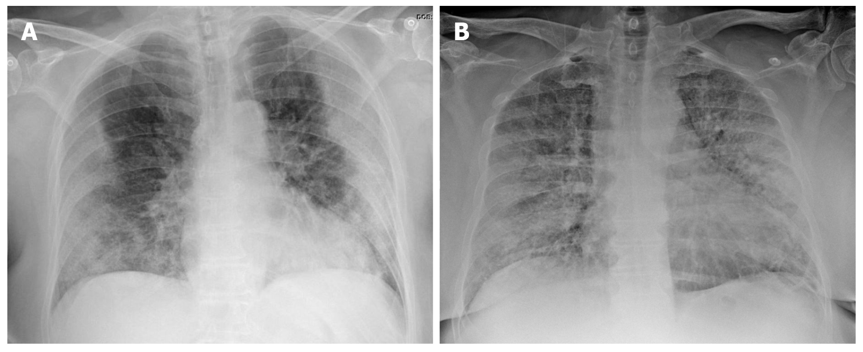
Figure 2 Chest X-ray findings in two patients with confirmed severe acute respiratory syndrome coronavirus 2 pneumonia (positive reverse transcription polymerase chain reaction test).
A: 73-year-old diabetic woman. General malaise, myalgia and diarrhea of 8 d clinical course. Dyspnea in the last 2 d, no fever. AP X-ray: Peripherally-distributed bilateral lung opacities; B: 60-year-old man, fever and cough, progressive dyspnea. The patient presented at the emergency department with acute respiratory failure requiring admission to intensive care. AP X-ray: Alveolar infiltrates and diffusely-distributed, bilateral ground-glass opacities.
- Citation: Landete P, Quezada Loaiza CA, Aldave-Orzaiz B, Muñiz SH, Maldonado A, Zamora E, Sam Cerna AC, del Cerro E, Alonso RC, Couñago F. Clinical features and radiological manifestations of COVID-19 disease. World J Radiol 2020; 12(11): 247-260
- URL: https://www.wjgnet.com/1949-8470/full/v12/i11/247.htm
- DOI: https://dx.doi.org/10.4329/wjr.v12.i11.247


