Copyright
©The Author(s) 2020.
World J Radiol. Oct 28, 2020; 12(10): 213-230
Published online Oct 28, 2020. doi: 10.4329/wjr.v12.i10.213
Published online Oct 28, 2020. doi: 10.4329/wjr.v12.i10.213
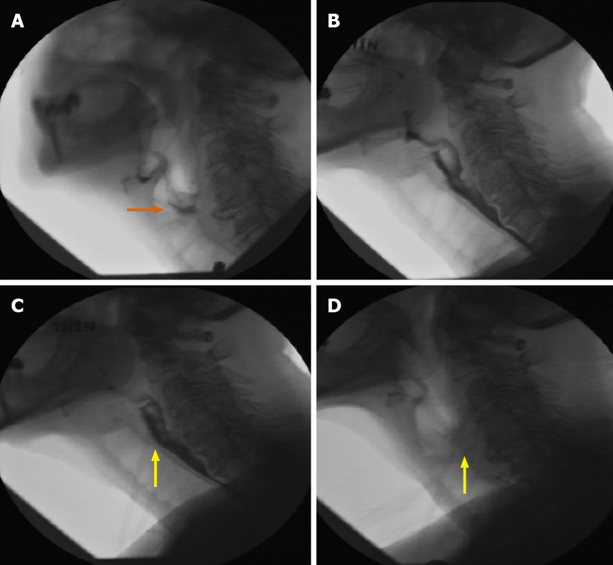
Figure 18 A 69-yr-old female.
A: Initial fluoroscopic examination of a patient with dysphagia demonstrating disorganized swallow and penetration to the level of the vocal cords (orange arrow); B: Follow-up fluoroscopic swallow examination (without neostigmine) demonstrating improvement in organized swallow with only mild flash penetration; C and D: Immediate follow-up examination with administration of neostigmine, showing no significant penetration at multiple consistencies, along with significant improvement in organized swallow (yellow arrows). Neurology consultation concurred with the diagnosis of myasthenia gravis, particularly based on the fluoroscopic series of examinations.
- Citation: Shalom NE, Gong GX, Auster M. Fluoroscopy: An essential diagnostic modality in the age of high-resolution cross-sectional imaging. World J Radiol 2020; 12(10): 213-230
- URL: https://www.wjgnet.com/1949-8470/full/v12/i10/213.htm
- DOI: https://dx.doi.org/10.4329/wjr.v12.i10.213


