Copyright
©The Author(s) 2020.
World J Radiol. Oct 28, 2020; 12(10): 213-230
Published online Oct 28, 2020. doi: 10.4329/wjr.v12.i10.213
Published online Oct 28, 2020. doi: 10.4329/wjr.v12.i10.213
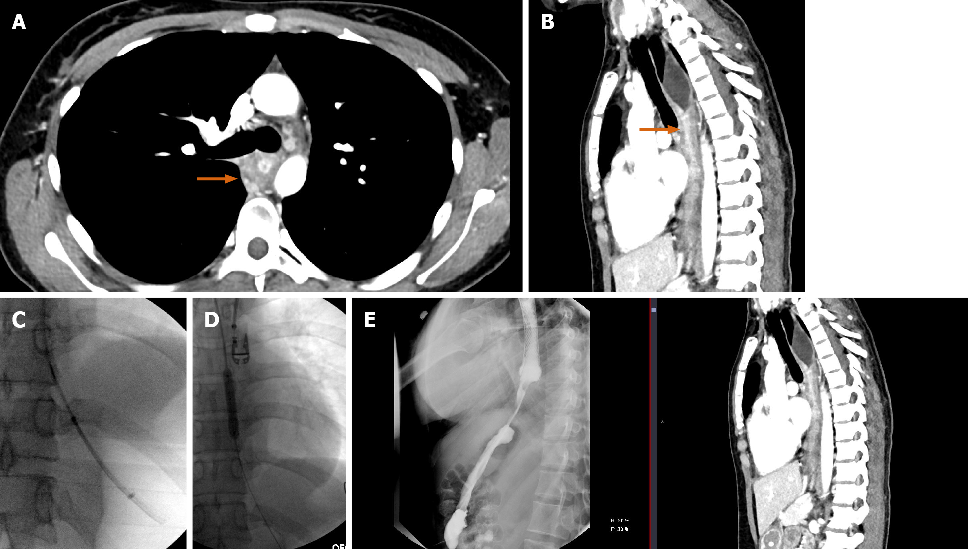
Figure 17 A 26-yr-old male.
A and B: Axial and sagittal computed tomography (CT) images showing long-segment esophageal stenosis related to caustic ingestion (orange arrows); C and D: With an initial upper endoscopy aborted, repeat upper endoscopy with the addition of fluoroscopic guidance allowed passage through a known stricture and eventual stent placement; E: Subsequent fluoroscopic upper gastrointestinal examination showing free contrast passage through the stented esophagus, with direct sagittal comparison to the initial CT in the sagittal plane.
- Citation: Shalom NE, Gong GX, Auster M. Fluoroscopy: An essential diagnostic modality in the age of high-resolution cross-sectional imaging. World J Radiol 2020; 12(10): 213-230
- URL: https://www.wjgnet.com/1949-8470/full/v12/i10/213.htm
- DOI: https://dx.doi.org/10.4329/wjr.v12.i10.213


