Copyright
©The Author(s) 2020.
World J Radiol. Oct 28, 2020; 12(10): 213-230
Published online Oct 28, 2020. doi: 10.4329/wjr.v12.i10.213
Published online Oct 28, 2020. doi: 10.4329/wjr.v12.i10.213
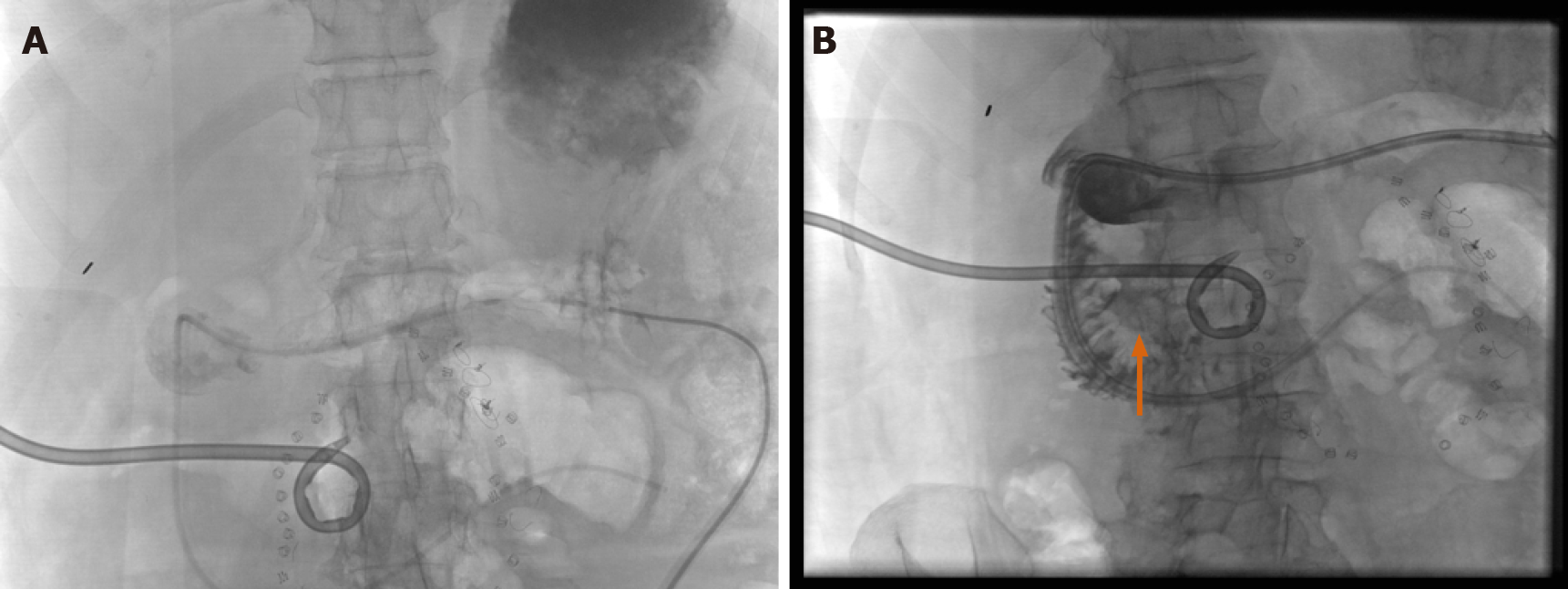
Figure 14 “Beyond computed tomography” in the determination of bowel extraluminal collections.
A: The supine position is utilized in computed tomography, where the G-port of a GJ tube was injected with contrast, then seen localizing to the dependent fundus; B: Real-time fluoroscopic projections with patient in the near-lateral position, showing a fistulous communication with anterior abscess and duodenal sweep (vertical orange arrow).
- Citation: Shalom NE, Gong GX, Auster M. Fluoroscopy: An essential diagnostic modality in the age of high-resolution cross-sectional imaging. World J Radiol 2020; 12(10): 213-230
- URL: https://www.wjgnet.com/1949-8470/full/v12/i10/213.htm
- DOI: https://dx.doi.org/10.4329/wjr.v12.i10.213


