Copyright
©The Author(s) 2020.
World J Radiol. Oct 28, 2020; 12(10): 213-230
Published online Oct 28, 2020. doi: 10.4329/wjr.v12.i10.213
Published online Oct 28, 2020. doi: 10.4329/wjr.v12.i10.213
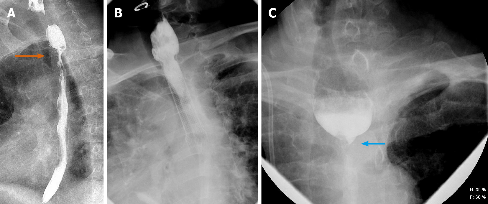
Figure 3 A 64-year-old male.
A-C: Carcinoma of the esophagus showing long segment moderate stenosis with irregular border and “apple core” appearance at the upper esophagus (orange arrow, upper left). Stent was placed, with follow-up exam showing worsening stricture, with near complete occlusion of the esophagus (blue arrow).
- Citation: Shalom NE, Gong GX, Auster M. Fluoroscopy: An essential diagnostic modality in the age of high-resolution cross-sectional imaging. World J Radiol 2020; 12(10): 213-230
- URL: https://www.wjgnet.com/1949-8470/full/v12/i10/213.htm
- DOI: https://dx.doi.org/10.4329/wjr.v12.i10.213


