Copyright
©The Author(s) 2019.
World J Radiol. Sep 28, 2019; 11(9): 116-125
Published online Sep 28, 2019. doi: 10.4329/wjr.v11.i9.116
Published online Sep 28, 2019. doi: 10.4329/wjr.v11.i9.116
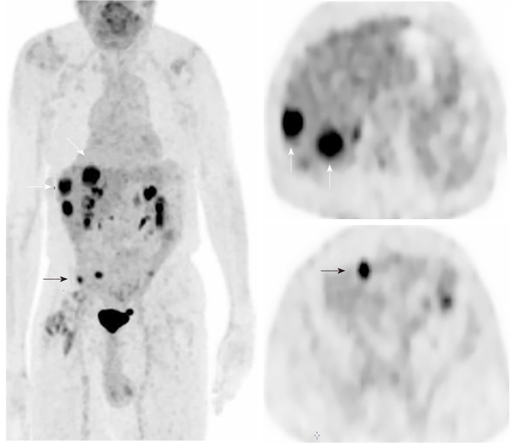
Figure 3 Coronal and axial fluorodeoxyglucose positron emission tomography images show fluorodexoyglucose avid liver (white arrows) and peritoneal (black arrows) metastases.
- Citation: Bhat AP, Schuchardt PA, Bhat R, Davis RM, Singh S. Metastatic appendiceal cancer treated with Yttrium 90 radioembolization and systemic chemotherapy: A case report. World J Radiol 2019; 11(9): 116-125
- URL: https://www.wjgnet.com/1949-8470/full/v11/i9/116.htm
- DOI: https://dx.doi.org/10.4329/wjr.v11.i9.116


