Copyright
©The Author(s) 2019.
World J Radiol. May 28, 2019; 11(5): 74-80
Published online May 28, 2019. doi: 10.4329/wjr.v11.i5.74
Published online May 28, 2019. doi: 10.4329/wjr.v11.i5.74
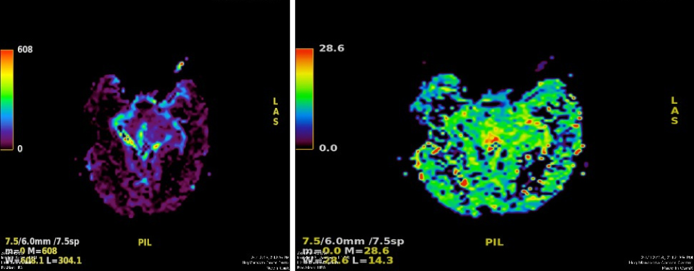
Figure 6 MR perfusion derived relative cerebral blood volume maps demonstrate heterogeneous perfusion abnormalities.
Mild higher normalized relative cerebral blood volume ratios in comparison with white matter are noted in the peripheral region of the cyst, which corresponds to malignant transformation.
- Citation: Pawar S, Borde C, Patil A, Nagarkar R. Malignant epidermoid arising from the third ventricle: A case report. World J Radiol 2019; 11(5): 74-80
- URL: https://www.wjgnet.com/1949-8470/full/v11/i5/74.htm
- DOI: https://dx.doi.org/10.4329/wjr.v11.i5.74


