Copyright
©The Author(s) 2019.
World J Radiol. Mar 28, 2019; 11(3): 27-45
Published online Mar 28, 2019. doi: 10.4329/wjr.v11.i3.27
Published online Mar 28, 2019. doi: 10.4329/wjr.v11.i3.27
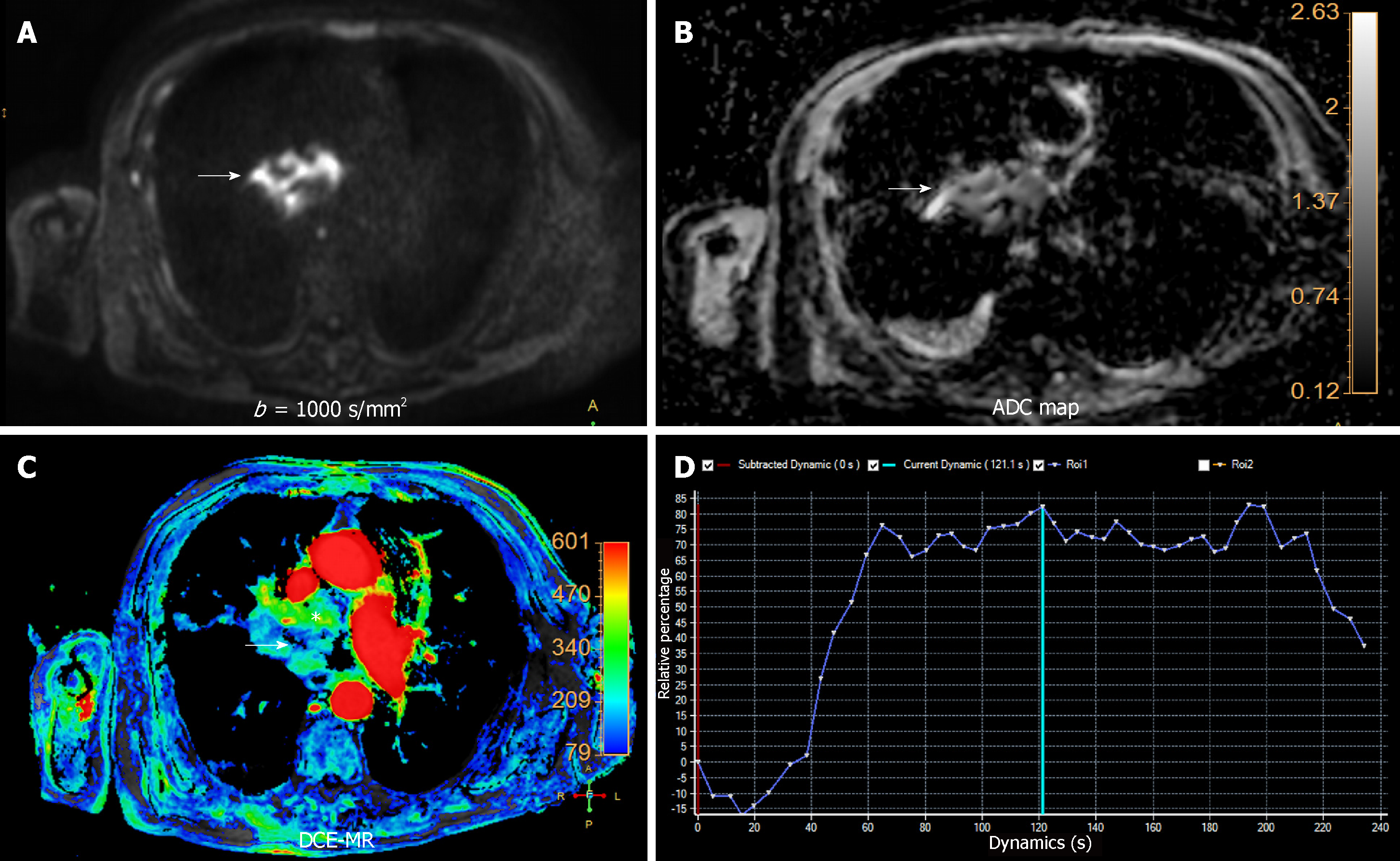
Figure 7 Multiparametric functional chest magnetic resonance of central lung cancer.
A 78 year-old male with a central hilar mass in keeping with epidermoid type lung carcinoma. A and B: High b value (b = 1000 s/mm2) and apparent diffusion coefficient (ADC) map (B) revealing a heterogeneous and restrictive behavior of the lesion, related to hypercellularity and aggressiveness (white arrows on A and B). The mean ADC was 0.97 × 10⁻³ mm2/s, confirming a malignant origin; C: Dynamic contrast-enhanced magnetic resonance showing a significant uptake of gadolinium based contrast agent on area under the curve parametric map (white asterisk), with central necrosis (White arrow on C); D: Time intensity curve (TIC) plot revealing steep slope of enhancement with posterior plateau (type B TIC) also favoring a malignant etiology. DCE-MR: Dynamic contrast-enhanced magnetic resonance; ADC: Apparent diffusion coefficient; TIC: Time intensity curve.
- Citation: Broncano J, Alvarado-Benavides AM, Bhalla S, Álvarez-Kindelan A, Raptis CA, Luna A. Role of advanced magnetic resonance imaging in the assessment of malignancies of the mediastinum. World J Radiol 2019; 11(3): 27-45
- URL: https://www.wjgnet.com/1949-8470/full/v11/i3/27.htm
- DOI: https://dx.doi.org/10.4329/wjr.v11.i3.27


