Copyright
©The Author(s) 2019.
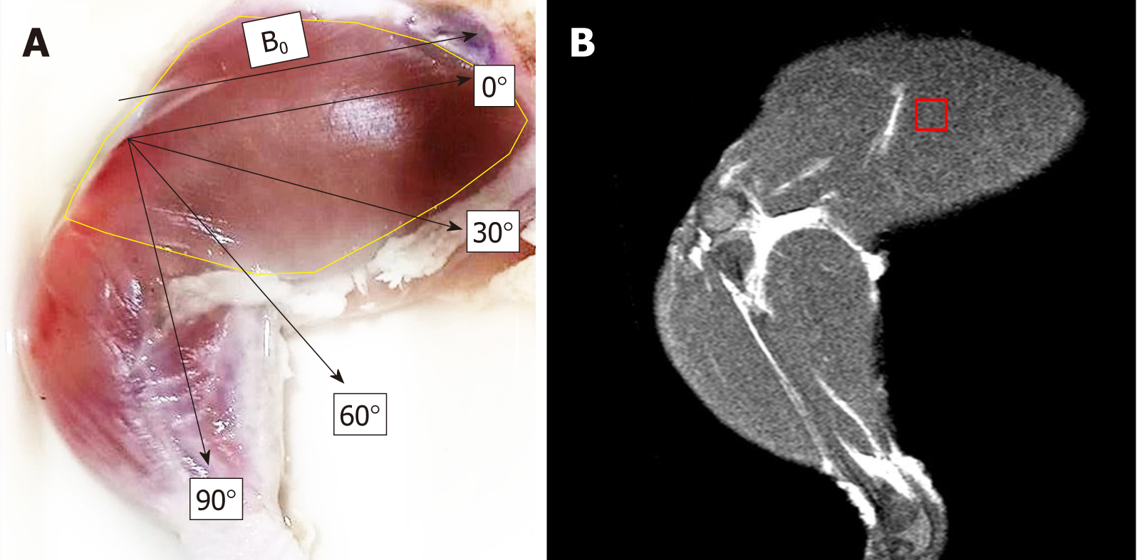
Figure 1 Chicken’s extensor iliotibialis was used to perform muscle quantification for muscle metabolites by proton magnetic resonance spectroscopy.
A: Skinned chicken thigh showing the extensor iliotibialis lateralis muscle fiber orientation (in the yellow marked area) at various angles (0°, 30°, 60°, and 90°) with regard to the main magnetic field (B0); B: T2-weighted turbo spin echo MRI images show areas of proton magnetic resonance spectroscopy voxel placement. B0: Main magnetic field direction.
- Citation: Pasanta D, Kongseha T, Kothan S. Effects of muscle fiber orientation to main magnetic field on muscle metabolite profiles for magnetic resonance spectroscopy acquisition. World J Radiol 2019; 11(1): 1-9
- URL: https://www.wjgnet.com/1949-8470/full/v11/i1/1.htm
- DOI: https://dx.doi.org/10.4329/wjr.v11.i1.1


