Copyright
©The Author(s) 2018.
World J Radiol. Sep 28, 2018; 10(9): 91-99
Published online Sep 28, 2018. doi: 10.4329/wjr.v10.i9.91
Published online Sep 28, 2018. doi: 10.4329/wjr.v10.i9.91
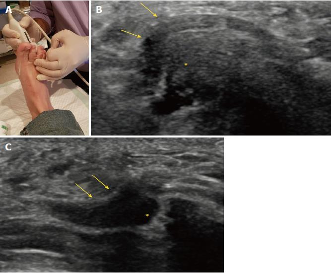
Figure 4 Displacement of Morton’s neuroma.
A: Pressure on the dorsal aspect of the web space; B, C: Long axis views of the neuroma and bursa before (B) and after (C) pressure of the dorsal aspect of the intermetatarsal space. In all images, arrows indicate Morton’s neuroma and asterisks (*) indicate bursa.
- Citation: Santiago FR, Muñoz PT, Pryest P, Martínez AM, Olleta NP. Role of imaging methods in diagnosis and treatment of Morton’s neuroma. World J Radiol 2018; 10(9): 91-99
- URL: https://www.wjgnet.com/1949-8470/full/v10/i9/91.htm
- DOI: https://dx.doi.org/10.4329/wjr.v10.i9.91


