Copyright
©The Author(s) 2018.
World J Radiol. Jul 28, 2018; 10(7): 78-82
Published online Jul 28, 2018. doi: 10.4329/wjr.v10.i7.78
Published online Jul 28, 2018. doi: 10.4329/wjr.v10.i7.78
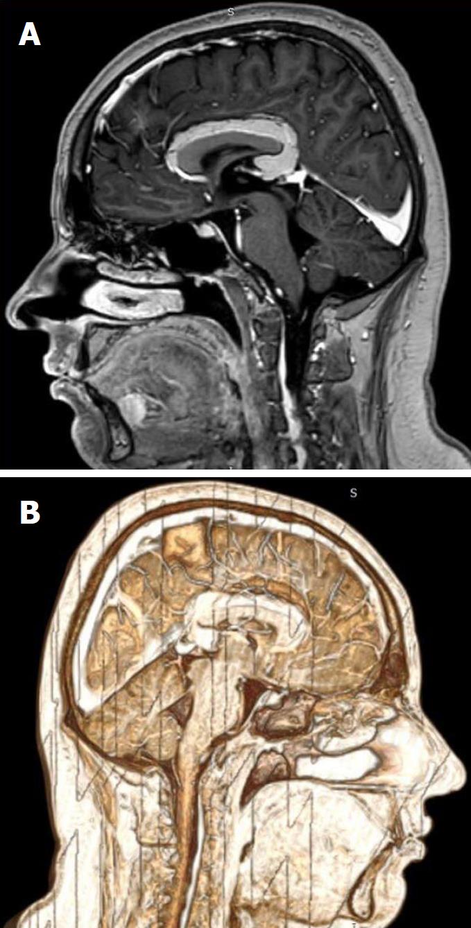
Figure 4 T1-W 3D TFE + MDC magnetic resonance imaging.
A: Showing the relationships between vasculature, corpus callosum, lipoma and adjacent brain tissues; B: Showing the pericallosal lesion, compatible with a complete curvilinear lipoma without callosal aplasia.
- Citation: Spanu F, Saba L. Obesity and pericallosal lipoma in X-linked emery-dreifuss muscular dystrophy: A case report - Does Emerin play a role in adipocyte differentiation? World J Radiol 2018; 10(7): 78-82
- URL: https://www.wjgnet.com/1949-8470/full/v10/i7/78.htm
- DOI: https://dx.doi.org/10.4329/wjr.v10.i7.78


