Copyright
©The Author(s) 2018.
World J Radiol. Jul 28, 2018; 10(7): 78-82
Published online Jul 28, 2018. doi: 10.4329/wjr.v10.i7.78
Published online Jul 28, 2018. doi: 10.4329/wjr.v10.i7.78
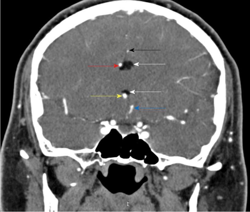
Figure 2 Coronal DSA.
Shows the lipoma (white arrows), the right rostral A2 (yellow arrow), the right pericallosal artery (red arrow), the left rostral A2 (blue arrow), and the left pericallosal artery (black arrow).
- Citation: Spanu F, Saba L. Obesity and pericallosal lipoma in X-linked emery-dreifuss muscular dystrophy: A case report - Does Emerin play a role in adipocyte differentiation? World J Radiol 2018; 10(7): 78-82
- URL: https://www.wjgnet.com/1949-8470/full/v10/i7/78.htm
- DOI: https://dx.doi.org/10.4329/wjr.v10.i7.78


