Copyright
©The Author(s) 2018.
World J Radiol. Jun 28, 2018; 10(6): 52-64
Published online Jun 28, 2018. doi: 10.4329/wjr.v10.i6.52
Published online Jun 28, 2018. doi: 10.4329/wjr.v10.i6.52
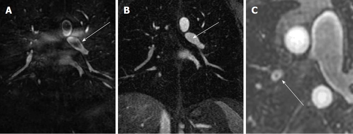
Figure 4 Artifacts: The Maki artifact.
A: Acquisition of the central aspect of k-space was before the bolus of contrast agent filled the pulmonary artery causing a pseudo-clot within the left lower lobe pulmonary artery (arrow); B: Later phase acquisition from the same patient shows normal contrast enhancement of the Left lower lobe pulmonary artery (arrow); C: Gibbs’’ ringing artifact can simulate a central filling defect. Typically, the signal of emboli will be less than 50% of the signal intensity of the lumen.
- Citation: Tsuchiya N, Beek EJV, Ohno Y, Hatabu H, Kauczor HU, Swift A, Vogel-Claussen J, Biederer J, Wild J, Wielpütz MO, Schiebler ML. Magnetic resonance angiography for the primary diagnosis of pulmonary embolism: A review from the international workshop for pulmonary functional imaging. World J Radiol 2018; 10(6): 52-64
- URL: https://www.wjgnet.com/1949-8470/full/v10/i6/52.htm
- DOI: https://dx.doi.org/10.4329/wjr.v10.i6.52


