Copyright
©The Author(s) 2018.
World J Radiol. Jun 28, 2018; 10(6): 52-64
Published online Jun 28, 2018. doi: 10.4329/wjr.v10.i6.52
Published online Jun 28, 2018. doi: 10.4329/wjr.v10.i6.52
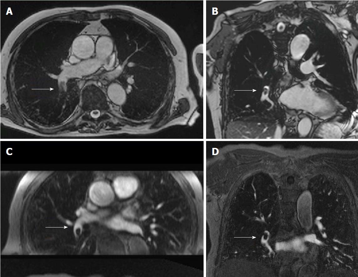
Figure 3 Non-contrast pulmonary magnetic resonance angiograph of an 82-year-old male with a history of san acute onset of dyspnea.
A: Transverse non-contrast enhanced steady state GRE; B: Coronal oblique non-contrast enhanced steady-state GRE images of a fresh embolus in the right lower lobe artery; C: For comparison the transverse reformation; D: The original coronal images from the contrast-enhanced MRA (images courtesy of Heussel CP and Wielpuetz M, Thoraxklinik, Heidelberg, Germany). GRE: Gradient recalled echo; MRA: Magnetic resonance angiograph.
- Citation: Tsuchiya N, Beek EJV, Ohno Y, Hatabu H, Kauczor HU, Swift A, Vogel-Claussen J, Biederer J, Wild J, Wielpütz MO, Schiebler ML. Magnetic resonance angiography for the primary diagnosis of pulmonary embolism: A review from the international workshop for pulmonary functional imaging. World J Radiol 2018; 10(6): 52-64
- URL: https://www.wjgnet.com/1949-8470/full/v10/i6/52.htm
- DOI: https://dx.doi.org/10.4329/wjr.v10.i6.52


