Copyright
©The Author(s) 2018.
World J Radiol. Jun 28, 2018; 10(6): 52-64
Published online Jun 28, 2018. doi: 10.4329/wjr.v10.i6.52
Published online Jun 28, 2018. doi: 10.4329/wjr.v10.i6.52
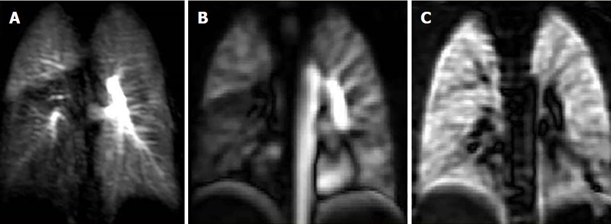
Figure 2 Case of pulmonary embolism to the right lower lobe.
A: Coronal dynamic contrast MRI shows notable right lower lobe hypo-perfusion in a 25-year-old female with known acute pulmonary embolism one month ago; B: Corresponding (non-contrast) Fourier decomposition (FD) perfusion; C: Ventilation-weighted FD MR images also depict right lower lobe hypo-perfusion and normal ventilation (VQ mismatch). MRI: Magnetic resonance imaging.
- Citation: Tsuchiya N, Beek EJV, Ohno Y, Hatabu H, Kauczor HU, Swift A, Vogel-Claussen J, Biederer J, Wild J, Wielpütz MO, Schiebler ML. Magnetic resonance angiography for the primary diagnosis of pulmonary embolism: A review from the international workshop for pulmonary functional imaging. World J Radiol 2018; 10(6): 52-64
- URL: https://www.wjgnet.com/1949-8470/full/v10/i6/52.htm
- DOI: https://dx.doi.org/10.4329/wjr.v10.i6.52


