Copyright
©The Author(s) 2018.
World J Radiol. May 28, 2018; 10(5): 46-51
Published online May 28, 2018. doi: 10.4329/wjr.v10.i5.46
Published online May 28, 2018. doi: 10.4329/wjr.v10.i5.46
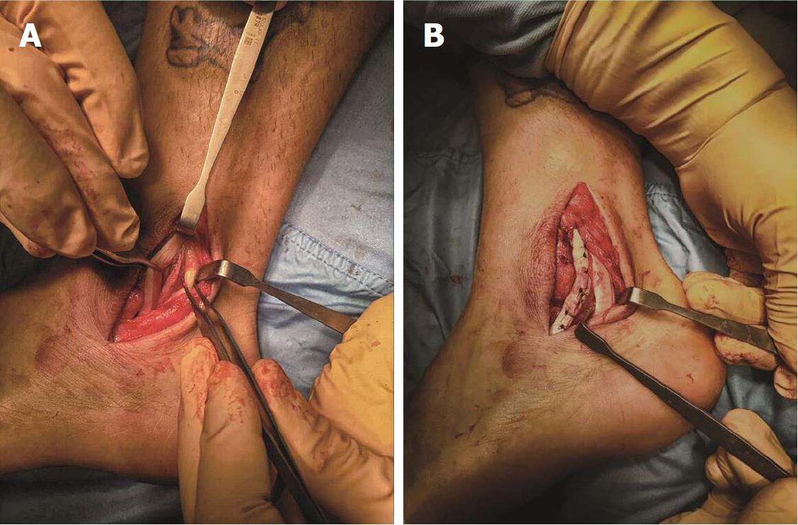
Figure 3 The surgical procedure.
A: Surgical view: The peroneus longus (PL) resides more posteriorly, while is well visible the peroneus brevis (PB) is torn in two hemi-tendons the anterior part is about the 70% of the total peroneus brevis tendon; B: At the end of operation the two PB hemi-tendons are sutured together; posteriorly PL can be appreciated.
- Citation: Fischetti A, Zawaideh JP, Orlandi D, Belfiore S, SIlvestri E. Traumatic peroneal split lesion with retinaculum avulsion: Diagnosis and post-operative multymodality imaging. World J Radiol 2018; 10(5): 46-51
- URL: https://www.wjgnet.com/1949-8470/full/v10/i5/46.htm
- DOI: https://dx.doi.org/10.4329/wjr.v10.i5.46


