Copyright
©The Author(s) 2018.
World J Radiol. May 28, 2018; 10(5): 46-51
Published online May 28, 2018. doi: 10.4329/wjr.v10.i5.46
Published online May 28, 2018. doi: 10.4329/wjr.v10.i5.46
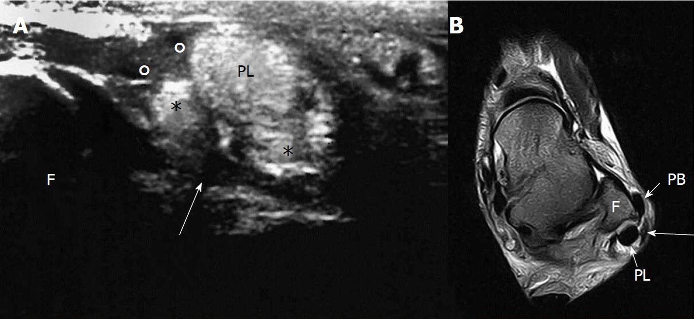
Figure 2 Ultrasound and magnetic resonance imaging evaluation of the injured ankle.
A: US examination of the injured ankle: Avulsed retinaculum (°); two hemi-tendons due to split lesion of peroneus brevis (*); CFL (arrow). B: MRI T2w axial sequence: MRI shows PB anterior subluxation. The avulsion of the retinaculum (arrow) can be observed. US: Ultrasound; MRI: Magnetic resonance imaging; PB: Peroneus brevis; PL: Peroneus longus.
- Citation: Fischetti A, Zawaideh JP, Orlandi D, Belfiore S, SIlvestri E. Traumatic peroneal split lesion with retinaculum avulsion: Diagnosis and post-operative multymodality imaging. World J Radiol 2018; 10(5): 46-51
- URL: https://www.wjgnet.com/1949-8470/full/v10/i5/46.htm
- DOI: https://dx.doi.org/10.4329/wjr.v10.i5.46


