Copyright
©The Author(s) 2018.
World J Radiol. May 28, 2018; 10(5): 46-51
Published online May 28, 2018. doi: 10.4329/wjr.v10.i5.46
Published online May 28, 2018. doi: 10.4329/wjr.v10.i5.46
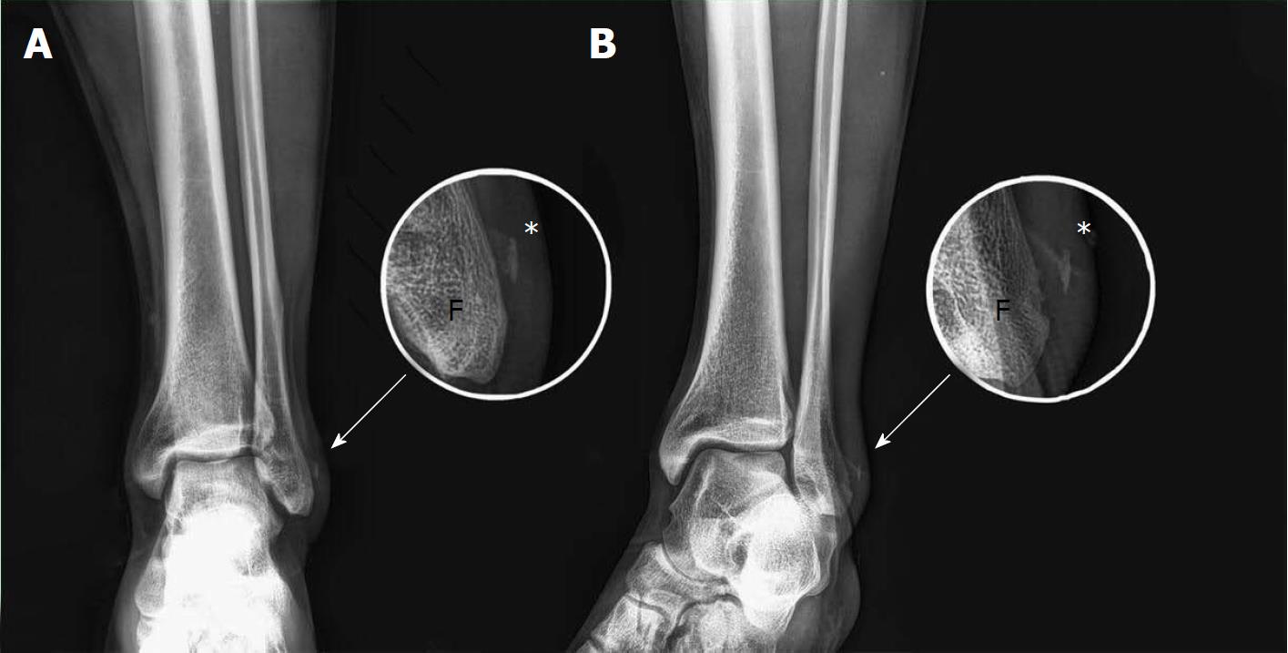
Figure 1 X-ray: AP and oblique view, no fractures of the peroneal malleolus are shown.
On the magnification a detachment of a small bony foil (periosteum) is appreciable (*). A: AP; B: Oblique view. F: Fibula.
- Citation: Fischetti A, Zawaideh JP, Orlandi D, Belfiore S, SIlvestri E. Traumatic peroneal split lesion with retinaculum avulsion: Diagnosis and post-operative multymodality imaging. World J Radiol 2018; 10(5): 46-51
- URL: https://www.wjgnet.com/1949-8470/full/v10/i5/46.htm
- DOI: https://dx.doi.org/10.4329/wjr.v10.i5.46


