Copyright
©The Author(s) 2018.
World J Radiol. Apr 28, 2018; 10(4): 30-45
Published online Apr 28, 2018. doi: 10.4329/wjr.v10.i4.30
Published online Apr 28, 2018. doi: 10.4329/wjr.v10.i4.30
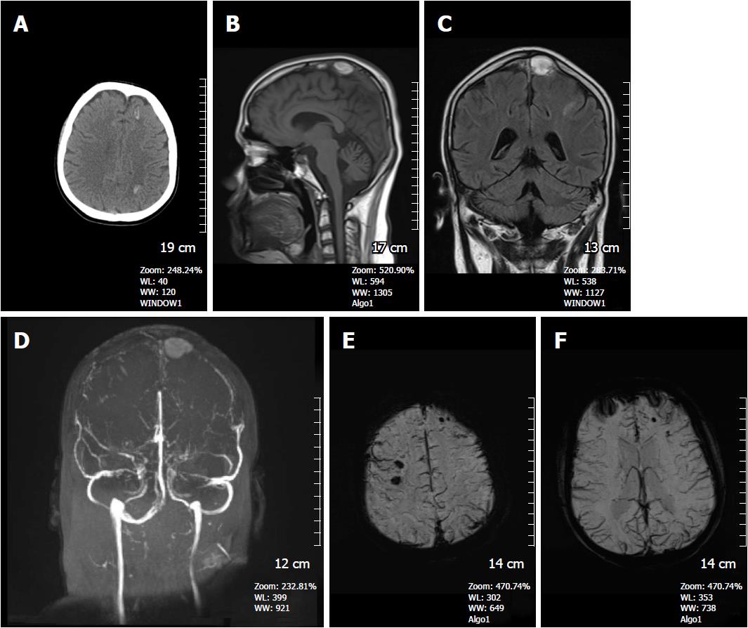
Figure 9 A 16-year-old man presented with superior sagittal sinus thrombosis.
A: Non-contrast CT image shows hyperdense hemorrhagic foci in the left frontal and left parietal lobe at the convexity level (arrows); B: On the sagittal T1W image, a hyperintense thrombus is detected in the superior sagittal sinus; C: Coronal FSE T2W image, thrombus again shows hyperintense signal intensity; D: Time of flight non-contrast MR venography, absence of normal venous flow and accompanying thrombus are clearly depicted; E and F: SWI minIP images demonstrates hypointense microhemorrhages in the brain parenchyma with diffuse dilated venous structures indicating venous engorgement due to venous hypertension. SWI: Susceptibility weighted imaging; minIP: Minimum intensity projection algorithm.
- Citation: Halefoglu AM, Yousem DM. Susceptibility weighted imaging: Clinical applications and future directions. World J Radiol 2018; 10(4): 30-45
- URL: https://www.wjgnet.com/1949-8470/full/v10/i4/30.htm
- DOI: https://dx.doi.org/10.4329/wjr.v10.i4.30


