Copyright
©The Author(s) 2018.
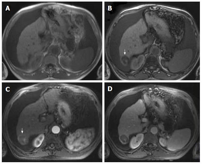
Figure 10 Fatty hepatocellular carcinoma.
Dual echo in-phase (A) and out-of-phase (B) GRE T1 weighted images and post-contrast fat-suppressed 3D-GRE T1-weighted images during late hepatic arterial (C) and delayed phase (D). A right hepatic lobe nodule shows loss of signal intensity on the out-of-phase (arrow, B) in comparison with the in-phase (A) images. This nodule shows heterogeneous arterial hyperenhancement (arrow, C) and a clear washout is appreciable (D) in keeping with fatty hepatocellular carcinoma.
- Citation: Campos-Correia D, Cruz J, Matos AP, Figueiredo F, Ramalho M. Magnetic resonance imaging ancillary features used in Liver Imaging Reporting and Data System: An illustrative review. World J Radiol 2018; 10(2): 9-23
- URL: https://www.wjgnet.com/1949-8470/full/v10/i2/9.htm
- DOI: https://dx.doi.org/10.4329/wjr.v10.i2.9


