Copyright
©The Author(s) 2018.
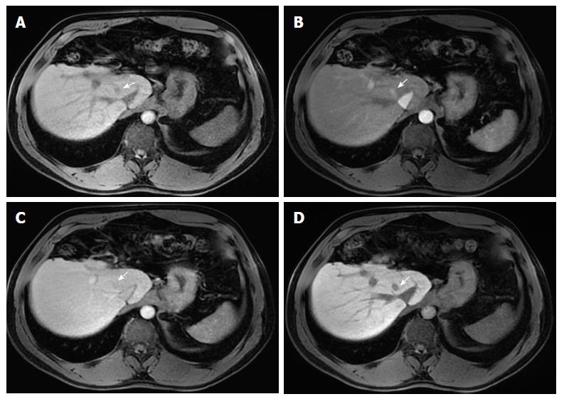
Figure 7 Small hepatocellular carcinoma in a patient with chronic liver disease and a previous left hepatectomy.
Pre- (A) and post-contrast (gadoxetate disodium) fat-suppressed 3D-GRE T1-weighted images in the arterial (B), delayed (C) and hepatobiliary phases (D). A small HCC is shown in segment VIII displaying mild low T1 signal intensity (arrow, A), arterial hyper-enhancement (arrow, B) and no appreciable washout on the delayed phase (arrow, C). Low signal intensity is noted on the hepatobiliary phase (arrow, D). This example proves the benefit of hepatobiliary contrast agents in further characterization of liver nodules in the setting of chronic liver disease.
- Citation: Campos-Correia D, Cruz J, Matos AP, Figueiredo F, Ramalho M. Magnetic resonance imaging ancillary features used in Liver Imaging Reporting and Data System: An illustrative review. World J Radiol 2018; 10(2): 9-23
- URL: https://www.wjgnet.com/1949-8470/full/v10/i2/9.htm
- DOI: https://dx.doi.org/10.4329/wjr.v10.i2.9


