Copyright
©The Author(s) 2018.
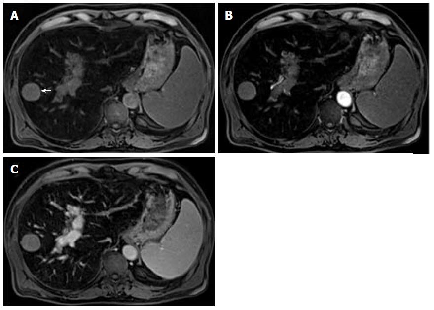
Figure 6 Lesional iron sparing.
Pre-contrast (A) and post-contrast fat-suppressed 3D-GRE T1-weighted images during the late hepatic arterial (B) and delayed (C) phases. An iron-overloaded cirrhotic liver shows marked signal loss on T1-weighted images. A non-siderotic nodule is seen in the right liver lobe (arrow, A). Dynamic evaluation is limited and does not allow a confident diagnosis of hepatocellular carcinoma (HCC). This nodule underwent biopsy and HCC was confirmed.
- Citation: Campos-Correia D, Cruz J, Matos AP, Figueiredo F, Ramalho M. Magnetic resonance imaging ancillary features used in Liver Imaging Reporting and Data System: An illustrative review. World J Radiol 2018; 10(2): 9-23
- URL: https://www.wjgnet.com/1949-8470/full/v10/i2/9.htm
- DOI: https://dx.doi.org/10.4329/wjr.v10.i2.9


