Copyright
©The Author(s) 2018.
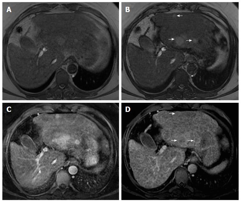
Figure 5 Lesional fat-sparing, in a case of multifocal hepatocellular carcinoma in a cirrhotic liver.
Dual echo in- (A) and out-of-phase (B) 2D-GRE T1 weighted images, post-contrast fat-suppressed 3D-GRE T1-weighted images during the late hepatic arterial (C) and delayed phase (D). In a patient with cirrhosis and fatty infiltration, perceived as loss of signal intensity in the out-of-phase images (B) in comparison with the in-phase images (A), three lesions with lower fractional fat content than the background liver are seen with higher signal intensity (arrows, B). These lesions show arterial hyperenhancement (C) and washout and late capsule enhancement in the delayed phase (arrows, D), consistent with HCCs.
- Citation: Campos-Correia D, Cruz J, Matos AP, Figueiredo F, Ramalho M. Magnetic resonance imaging ancillary features used in Liver Imaging Reporting and Data System: An illustrative review. World J Radiol 2018; 10(2): 9-23
- URL: https://www.wjgnet.com/1949-8470/full/v10/i2/9.htm
- DOI: https://dx.doi.org/10.4329/wjr.v10.i2.9


